. 2010 Sep 9;5(9):e12624.
doi: 10.1371/journal.pone.0012624.

Connexin mediated cataract prevention in mice

Affiliations

Connexin mediated cataract prevention in mice

Lin Li et al. PLoS One. .

Abstract

Cataracts, named for any opacity in the ocular lens, remain the leading cause of vision loss in the world. Non-surgical methods for cataract prevention are still elusive. We have genetically tested whether enhanced lens gap junction communication, provided by increased α3 connexin (Cx46) proteins expressed from α8(Kiα3) knock-in alleles in Gja8tm1(Gja3)Tww mice, could prevent nuclear cataracts caused by the γB-crystallin S11R mutation in CrygbS11R/S11R mice. Remarkably, homozygous knock-in α8(Kiα3/Kiα3) mice fully prevented nuclear cataracts, while single knock-in α8(Kiα3/-) allele mice showed variable suppression of nuclear opacities in CrygbS11R/S11R mutant mice. Cataract prevention was correlated with the suppression of many pathological processes, including crystallin degradation and fiber cell degeneration, as well as preservation of normal calcium levels and stable actin filaments in the lens. This work demonstrates that enhanced intercellular gap junction communication can effectively prevent or delay nuclear cataract formation and suggests that small metabolites transported through gap junction channels protect the stability of crystallin proteins and the cytoskeletal structures in the lens core. Thus, the use of an array of small molecules to promote lens homeostasis may become a feasible non-surgical approach for nuclear cataract prevention in the future.


Conflict of interest statement

Competing Interests: The authors have declared that no competing interests exist.

Figures

Figure 1
Figure 1. Cataract prevention mediated by α3 connexin in the γB-S11R mutation.
(A) Photos of wild-type (WT), mutant γB(S11R/S11R) and compound mutant γB(S11R/S11R) α8(Kiα3/Kiα3) lenses from P21 mice. Scale bar, 1 mm. (B) Representative light scattering graphs, obtained by an optical fiber and spectrometer, from P21 WT (blue), γB(S11R/S11R) (red) and γB(S11R/S11R) α8(Kiα3/Kiα3) (green) lenses. (C) The bar graph compares the normalized intensity of scattered light (y-axis with arbitrary units), calculated from the averaged area below the light scattering peaks of WT, γB(S11R/S11R) and γB(S11R/S11R) α8(Kiα3/Kiα3) lenses (x-axis).
Figure 2
Figure 2. Histology of P7 WT, γB(S11R/S11R) and γB(S11R/S11R) α8(Kiα3/Kiα3) lenses.
Wide field view of lens sections are shown on the left panels. Scale bar, 100 µm. High magnification views of selective interior fiber cells of lens sections are shown on the middle panels (dashed boxes, about 500–580 µm distance from the lens capsule) and the right panels (solid boxes, about 800–880 µm from the lens capsule). Uniformly elongated and tightly packed interior fiber cells are present in wild-type lens sections (the top panels) while irregularly elongated and loosely packed interior fiber cells (indicated by arrowheads on the middle panel) and disintegrated fiber cells (indicated by arrows on the middle-right panel) appears in γB(S11R/S11R) lenses. However, γB(S11R/S11R) α8(Kiα3/Kiα3) lens section displays uniformly elongated and tightly packed interior fiber cells without noticeable disintegrated fiber cells in the lens core (the bottom panels). Scale bars, 20 µm.
Figure 3
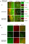
Figure 3. The distribution of actin filaments and cytosolic γ-crystallin in lens fiber cells.
(A) Postnatal day 1 (P1) WT, γB(S11R/S11R) and γB(S11R/S11R) α8(Kiα3/Kiα3) lens frozen sections were stained with anti-γ-crystallin antibody (green) and rhodamine-phalloidin (red). Select areas (boxes, about 460–530 µm from lens capsule) reveal both separated and merged fluorescent images of γ-crystallin and F-actin in lens inner fiber cells. Wild-type lenses show typical F-actin staining and uniformly distributed γ-crystallins while γB(S11R/S11R) lenses show both aberrant F-actin (arrowheads) and γ-crystallin aggregates along cell-cell boundaries (without F-actin, an arrow) in lens inner regions. Remarkably, γB(S11R/S11R) α8(Kiα3/Kiα3) lenses display normal F-actin with cytosolic γ-crystallins in inner fiber cells. Scale bars, 50 µm. (B) Enlarged view and merged fluorescent images of immunostained α3-connexin (green) with rhodamine-phalloidin-stained F-actin (red) in inner fiber cells of WT, γB(S11R/S11R) and γB(S11R/S11R) α8(Kiα3/Kiα3) lenses from P7 mice. Scale bars, 50 µm.
Figure 4
Figure 4. Levels of lens connexins and total lens calcium.
(A) Western blotting images and densitometric quantifications of the expression level of α3 connexin and α8 connexin in P10 WT, γB(S11R/S11R) and γB(S11R/S11R) α8(Kiα3/Kiα3) lenses. (B) Total lens calcium levels in P10 WT, γB(S11R/S11R) and γB(S11R/S11R) α8(Kiα3/Kiα3) lenses. (C) Western blotting images of lens total αA-, αB-, β-, γ-crystallin and actin. Arrowheads indicate the cleaved forms of crystallins. Two major cleaved forms of αA- and αB-crystallins are marked as αA-C1, αA-C2, αB-C1 and αB-C2, respectively.
Figure 5
Figure 5. Correlation between cleaved crystallin proteins and the severity of nuclear cataracts.
(A) Lens photos from three γB(S11R/S11R) α8(Kiα3/−) α3(−/−) littermates at the age of one month. Scale bar, 1 mm. (B) Western blotting images of lens total αA-, αB-, β- and γ-crystallins. Arrowheads indicate the cleaved forms of crystallins. Two major cleaved forms of αA- and αB-crystallins are marked as αA-C1, αA-C2, αB-C1, and αB-C2, respectively.

Similar articles

Cited by

References

    1. Asbell PA, Dualan I, Mindel J, Brocks D, Ahmad M, et al. Age-related cataract. Lancet. 2005;365:599–609. - PubMed
    1. Bloemendal H, de Jong W, Jaenicke R, Lubsen NH, Slingsby C, et al. Ageing and vision: structure, stability and function of lens crystallins. Prog Biophys Mol Biol. 2004;86:407–485. - PubMed
    1. Kuszak JR, Zoltoski RK, Sivertson C. Fibre cell organization in crystalline lenses. Exp Eye Res. 2004;78:673–687. - PubMed
    1. Nowak RB, Fischer RS, Zoltoski RK, Kuszak JR, Fowler VM. Tropomodulin1 is required for membrane skeleton organization and hexagonal geometry of fiber cells in the mouse lens. J Cell Biol. 2009;186:915–928. - PMC - PubMed
    1. Mathias RT, White TW, Gong X. Lens gap junctions in growth, differentiation, and homeostasis. Physiol Rev. 2010;90:179–206. - PMC - PubMed

Publication types