. 2010 Jan 1:2010:584657.
doi: 10.1155/2010/584657.

Role for PKC δ in Fenretinide-Mediated Apoptosis in Lymphoid Leukemia Cells

Affiliations

Role for PKC δ in Fenretinide-Mediated Apoptosis in Lymphoid Leukemia Cells

Vivian R Ruvolo et al. J Signal Transduct. .

Abstract

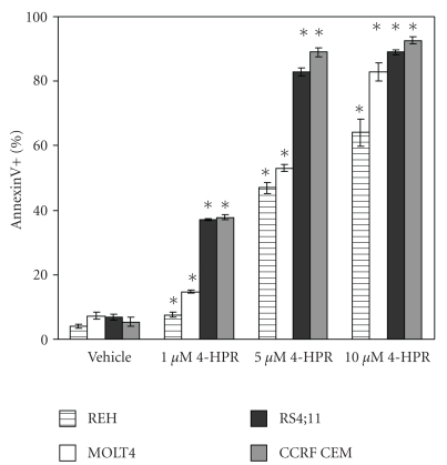
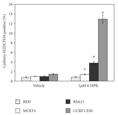
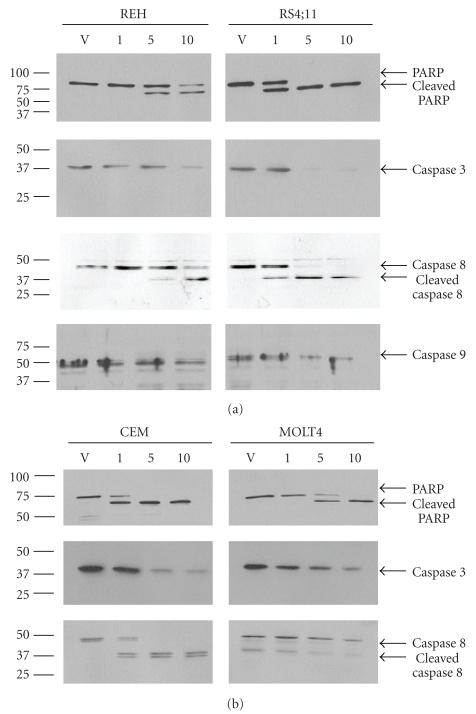
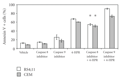
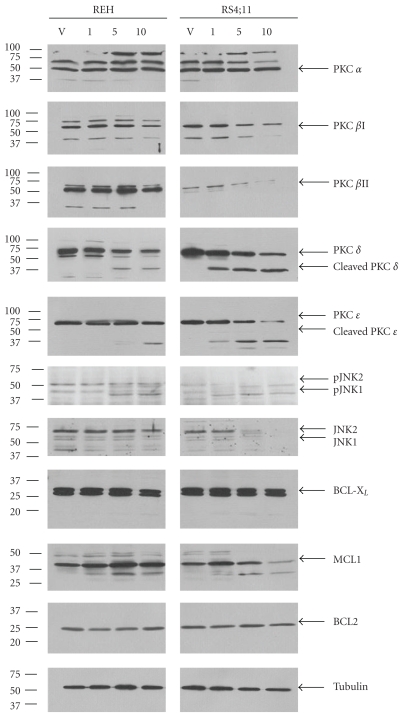
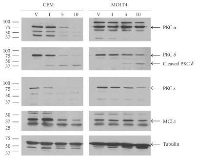
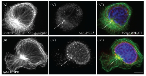
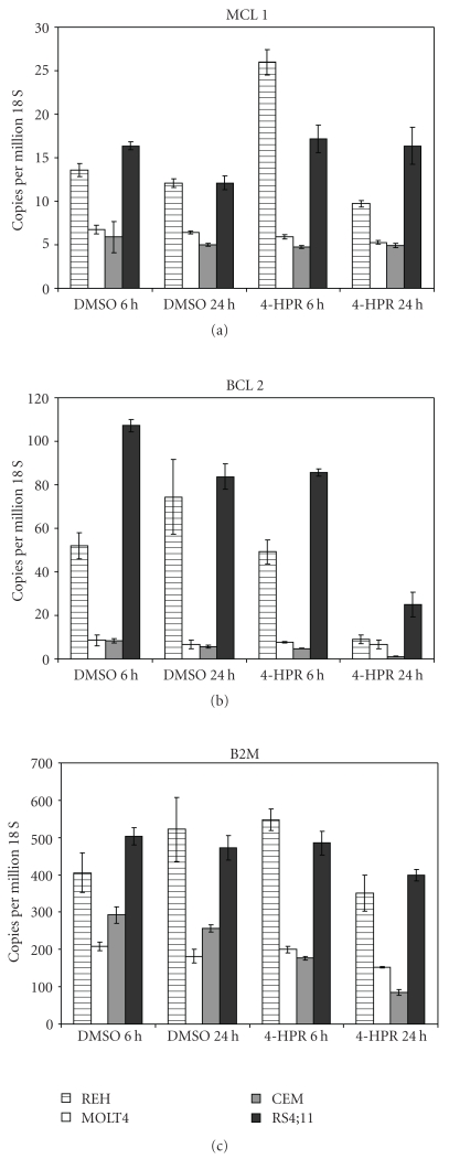
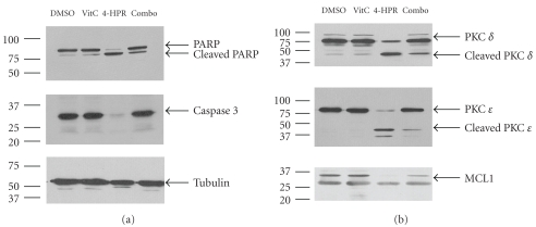
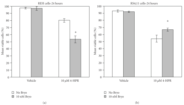
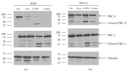
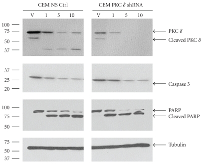
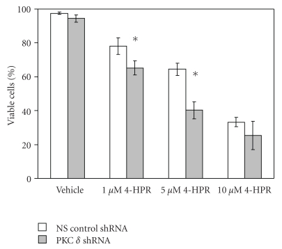
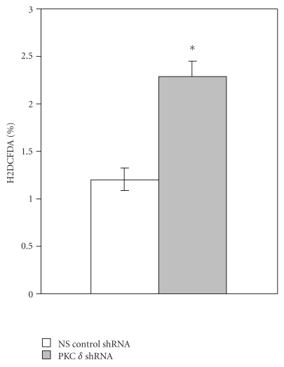
The synthetic Vitamin A analog fenretinide is a promising chemotherapeutic agent. In the current paper, the role of PKC δ was examined in fenretinide-induced apoptosis in lymphoid leukemia cells. Levels of proapoptotic cleaved PKC δ positively correlated with drug sensitivity. Fenretinide promoted reactive oxygen species (ROS) generation. The antioxidant Vitamin C prevented fenretinide-induced PKC δ cleavage and protected cells from fenretinide. Suppression of PKC δ expression by shRNA sensitized cells to fenretinide-induced apoptosis possibly by a mechanism involving ROS production. A previous study demonstrated that fenretinide promotes degradation of antiapoptotic MCL-1 in ALL cells via JNK. Now we have found that fenretinide-induced MCL-1 degradation may involve PKC δ as cleavage of the kinase correlated with loss of MCL-1 even in cells when JNK was not activated. These results suggest that PKC δ may play a complex role in fenretinide-induced apoptosis and may be targeted in antileukemia strategies that utilize fenretinide.


Figures

Figure 1
Figure 1
Fenretinide promotes apoptosis in leukemia cell lines. Apoptosis of human leukemia derived REH, MOLT4, RS4;11 and CCRF-CEM cells treated with vehicle (0.1% DMSO) or fenretinide (4-HPR at 1 μM, 5 μM or 10 μM dose) for 24 hours was examined using FACSCAN analysis of Annexin V stained cells. Error bars represent the mean ± S.D. from three separate experiments. Statistically significant differences from cell viability in untreated cells (standard t-test; P < .05) are marked by “∗”.
Figure 2
Figure 2
Fenretinide promotes ROS generation in leukemia cell lines. Generation of ROS in human leukemia derived REH, MOLT4, RS4;11 and CCRF-CEM cells treated with vehicle (0.1% DMSO) or fenretinide (1 μM 4-HPR) for 24 hours was examined using FACSCAN analysis of Carboxy-H2DCFDA stained cells. Error bars represent the mean ± S.D. from three separate experiments. Statistically significant differences from cell viability in untreated cells (standard t-test; P < .05) are marked by “∗”.
Figure 3
Figure 3
The antioxidant Vitamin C protects leukemia cell lines from fenretinide-induced apoptosis. Apoptosis of human leukemia derived REH, MOLT4, RS4;11 and CCRF-CEM cells treated with vehicle (0.2% DMSO) or fenretinide (10 μM 4-HPR) for 24 hours was examined using FACSCAN analysis of Annexin V stained cells. Where appropriate, cells were pretreated for 2 hours with 0.4 M Vitamin C (Vit C). Error bars represent the mean ± S.D. from three separate experiments. Statistically significant differences from cell viability in untreated cells (standard t-test; P < .05) are marked by “∗”.
Figure 4
Figure 4
Fenretinide promotes Caspase activity in leukemia cell lines. Western blot analysis was performed using antibody against PARP, Caspase 3, Caspase 8, and Caspase 9 on total lysate (0.25 × 10⁶ cell equivalents) from REH, RS4;11, MOLT4, and CCRF-CEM (CEM) cells treated with vehicle (lane marked V; 0.1% DMSO), 1 μM fenretinide (lane marked 1), 5 μM fenretinide (lane marked 5), or 10 μM fenretinide (lane marked 10).
Figure 5
Figure 5
Caspase Inhibitors do not protect leukemia cell lines from fenretinide-induced apoptosis. Apoptosis of human leukemia derived RS4;11 and CCRF-CEM (CEM) cells treated with vehicle (0.2% DMSO) or fenretinide (10 μM 4-HPR) for 24 hours was examined using FACSCAN analysis of Annexin V stained cells. Where appropriate, cells were pretreated for 2 hours with 20 μM Caspase 8 inhibitor or 20 μM Caspase 9 inhibitor. Error bars represent the mean ± S.D. from three separate experiments. Statistically significant differences from cell viability in untreated cells (standard t-test; P < .05) are marked by “∗”.
Figure 6
Figure 6
Fenretinide promotes PKC δ cleavage and suppresses MCL-1 expression in RS4;11 cells. Western blot analysis was performed using antibody against PKC α, PKC βI, PKC βII, PKC δ, PKC ε, p-JNK, JNK, BCL-XL, MCL-1, BCL2, and Tubulin on total lysate (0.25 × 10⁶ cell equivalents) from REH and RS4;11 cells treated with vehicle (lane marked V; 0.1% DMSO), 1 μM fenretinide (lane marked 1), 5 μM fenretinide (lane marked 5), or 10 μM fenretinide (lane marked 10) for 24 hours.
Figure 7
Figure 7
Fenretinide promotes PKC δ cleavage and suppresses MCL-1 expression in CCRF-CEM cells. Western blot analysis was performed using antibody against PKC α, PKC δ, PKC ε, MCL-1, and Tubulin on total lysate (0.25 × 10⁶ cell equivalents) from CCRF-CEM (CEM) cells and RS4;11 cells treated with vehicle (lane marked V; 0.1% DMSO), 1 μM fenretinide (lane marked 1), 5 μM fenretinide (lane marked 5), or 10 μM fenretinide (lane marked 10) for 24 hours.
Figure 8
Figure 8
PKC-δ translocates into the nucleus in response to fenretinide (4-HPR) treatment. CCRF-CEM cells were treated with either vehicle (A-A′′) or 1 μM 4-HPR (B-B′′) for 24 hours and then sedimented onto poly L-lysine coated coverslips. Cells were stained with anti α tubulin to label the microtubule network (A-B), and anti-PKC δ (A′-B′). In the untreated cells, the PKC δ localizes to the cytoplasm and is concentrated at the centrosome (A′, arrow). In the treated cells, the PKC δ is concentrated in the nucleus, and also at the cytoplasm (B′, arrow). Fluorescence optics Bar = 5 μM.
Figure 9
Figure 9
Fenretinide does not inhibit transcription of MCL-1 in leukemia cells. Real-Time-PCR was performed using cDNA derived from cells treated with vehicle (0.1% DMSO) or cells treated with 10 μM fenretinide (4-HPR) for 6 hours or 24 hours. Expression of MCL-1, BCL2, and B2M genes are presented as relative to 106 copies of 18S RNA.
Figure 10
Figure 10
The antioxidant Vitamin C blocks fenretinide-induced cleavage of PKC δ in RS4;11 cells. Western blot analysis was performed using antibody against PKC α, PKC βI, PKC βII, PKC δ, PKC ε, p-JNK, JNK, BCL-XL, MCL-1, BCL2, and Tubulin on total lysate (0.25 × 10⁶ cell equivalents) from RS4;11 cells treated with vehicle (0.2% DMSO), 0.4 M Vitamin C (Vit C), 10 μM fenretinide (4-HPR), or 10 μM fenretinide after a 2 hour pretreatment with 0.4 M Vitamin C (Combo) for 24 hours.
Figure 11
Figure 11
Bryostatin-1 protects RS4;11 but not REH cells from fenretinide-induced cell death. Cell death of human leukemia derived REH cells (a) and RS4;11 cells (b) treated with vehicle (0.2% DMSO) or fenretinide (10 μM 4-HPR) for 24 hours was examined by trypan blue dye exclusion assay. Where appropriate, cells were pretreated for 2 hours with 10 nM Bryostatin-1 (Bryo). Error bars represent the mean ± S.D. from three separate experiments. Statistically significant differences from cell viability in untreated cells (standard t-test; P < .05) are marked by “∗”.
Figure 12
Figure 12
Bryostatin-1 suppresses PKC δ expression in REH cells. Western blot analysis was performed using antibody against PKC δ, PKC ε, and Tubulin on total lysate (0.25 × 10⁶ cell equivalents) from REH cells and RS4;11 cells treated with vehicle (0.2% DMSO), 10 nM Bryostatin-1 (Bryo), fenretinide 4-HPR), or fenretinide after a 2 hour pretreatment with 10 nM Bryostatin-1 (Combo) for 24 hours. Due to differences in sensitivity to the drug, 10 μM fenretinide was used for REH cells and 1 μM fenretinide was used for RS4;11 cells.
Figure 13
Figure 13
Suppression of PKC δ promotes PARP cleavage in CCRF-CEM cells. Western blot analysis was performed using antibody against PKC δ, Caspase 3, PARP, and Tubulin on total lysate (0.25 × 10⁶ cell equivalents) from CCRF-CEM transfectant cells with control shRNA or CCRF-CEM transfectant cells with PKC δ shRNA that were treated with vehicle (lane marked V; 0.1% DMSO), 1 μM fenretinide (lane marked 1), 5 μM fenretinide (lane marked 5), or 10 μM fenretinide (lane marked 10) for 24 hours.
Figure 14
Figure 14
Suppression of PKC δ promotes fenretinide-induced cell death in CCRF-CEM cells. Cell death of CCRF-CEM transfectant cells with control nonspecific (NS) shRNA and CCRF-CEM transfectant cells with PKC δ (PKC delta) shRNA treated with vehicle (0.1% DMSO) or fenretinide (4-HPR at 1 μM, 5 μM or 10 μM dose) for 24 hours was examined by trypan blue dye exclusion assay. Error bars represent the mean ± S.D. from three separate experiments. Statistically significant differences from cell viability in untreated cells (standard t-test; P < .05) are marked by “∗”.
Figure 15
Figure 15
Suppression of PKC δ promotes ROS generation in CCRF-CEM. Generation of ROS in CCRF-CEM transfectant cells with control nonspecific (NS) shRNA and CCRF-CEM transfectant cells with PKC δ (PKC delta) shRNA was examined using FACSCAN analysis of Carboxy-H2DCFDA stained cells. Error bars represent the mean ± S.D. from three separate experiments. Statistically significant differences from cell viability in untreated cells (standard t-test; P < .05) are marked by “∗”.

Similar articles

Cited by

References

    1. Corazzari M, Lovat PE, Oliverio S, et al. Fenretinide: a p53-independent way to kill cancer cells. Biochemical and Biophysical Research Communications. 2005;331(3):810–815. - PubMed
    1. Lotan R. Retinoids and apoptosis: implications for cancer chemoprevention and therapy. Journal of the National Cancer Institute. 1995;87(22):1655–1657. - PubMed
    1. Maurer BJ, Melton L, Billups C, Cabot MC, Reynolds CP. Synergistic cytotoxicity in solid tumor cell lines between N-(4-hydroxyphenyl)retinamide and modulators of ceramide metabolism. Journal of the National Cancer Institute. 2000;92(23):1897–1909. - PubMed
    1. Sun S-Y, Li W, Yue P, Lippman SM, Hong WK, Lotan R. Mediation of N-(4-hydoxyphenyl)retinamide-induced apoptosis in human cancer cells by different mechanisms. Cancer Research. 1999;59(10):2493–2498. - PubMed
    1. O’Donnell PH, Guo W-X, Reynolds CP, Maurer BJ. N-(4-hydroxyphenyl)retinamide increases ceramide and is cytotoxic to acute lymphoblastic leukemia cell lines, but not to non-malignant lymphocytes. Leukemia. 2002;16(5):902–910. - PubMed