. 2010 Sep 14;1(4):e00171-10.
doi: 10.1128/mBio.00171-10.

Dendritic cell-specific antigen delivery by coronavirus vaccine vectors induces long-lasting protective antiviral and antitumor immunity

Affiliations

Dendritic cell-specific antigen delivery by coronavirus vaccine vectors induces long-lasting protective antiviral and antitumor immunity

Luisa Cervantes-Barragan et al. mBio. .

Abstract

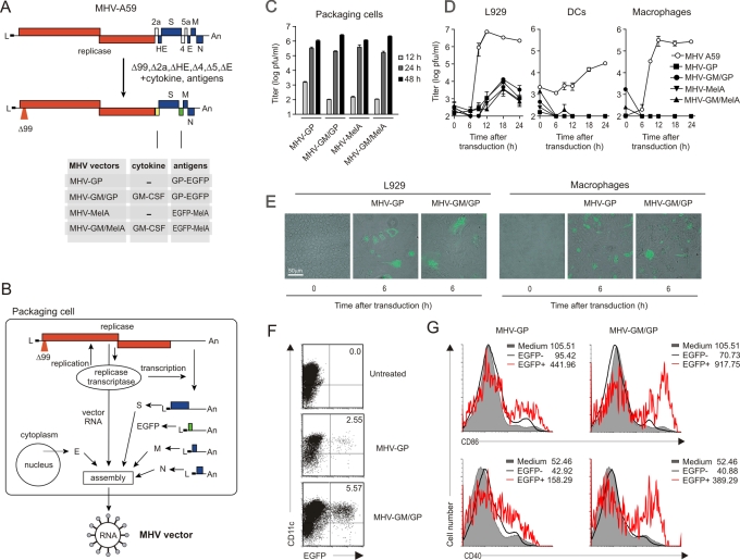
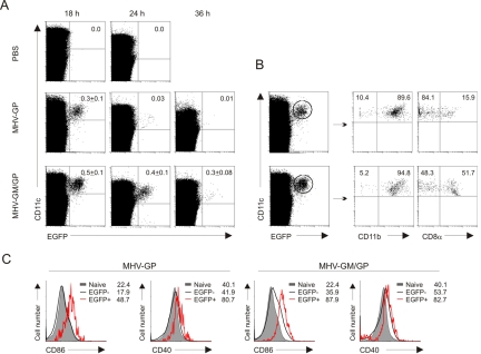
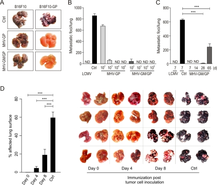
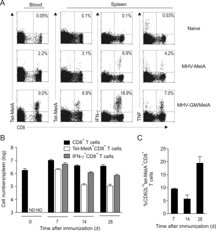
Efficient vaccination against infectious agents and tumors depends on specific antigen targeting to dendritic cells (DCs). We report here that biosafe coronavirus-based vaccine vectors facilitate delivery of multiple antigens and immunostimulatory cytokines to professional antigen-presenting cells in vitro and in vivo. Vaccine vectors based on heavily attenuated murine coronavirus genomes were generated to express epitopes from the lymphocytic choriomeningitis virus glycoprotein, or human Melan-A, in combination with the immunostimulatory cytokine granulocyte-macrophage colony-stimulating factor (GM-CSF). These vectors selectively targeted DCs in vitro and in vivo resulting in vector-mediated antigen expression and efficient maturation of DCs. Single application of only low vector doses elicited strong and long-lasting cytotoxic T-cell responses, providing protective antiviral and antitumor immunity. Furthermore, human DCs transduced with Melan-A-recombinant human coronavirus 229E efficiently activated tumor-specific CD8(+) T cells. Taken together, this novel vaccine platform is well suited to deliver antigens and immunostimulatory cytokines to DCs and to initiate and maintain protective immunity.


Figures

FIG 1
FIG 1
Generation, propagation, and in vitro target cell tropism of MHV-based vaccine vectors. (A) Schematic representation of the MHV-A59 genome and the highly attenuated MHV vectors. (B) Packaging concept for the generation of replication-competent but propagation-deficient MHV particles. (C) Growth kinetics of the indicated MHV vectors in 17ECl20 packaging cells. Cells were infected at an MOI of 1, and titers in supernatants were determined at the indicated time points (means of results from triplicate measurements ± standard errors of the means [SEM]). (D) Replication of the indicated MHV vectors or MHV-A59 wild-type virus in L929 cells, bone marrow-derived DCs, and peritoneal macrophages. The indicated cells (106/ml, MOI = 1) were infected, and replication was monitored by titration of supernatants on 17ECl20 packaging cells (mean ± SEM of duplicate measurements). One representative experiment out of three is displayed. (E) Transduction of L929 cells and peritoneal macrophages with an MHV-GP or MHV-GM/GP vector (MOI = 1). Green fluorescence was recorded 6 h posttransduction. Original magnification, ×400. (F, G) Stimulation of DCs by GM-CSF-expressing vectors. Bone marrow-derived DCs (106) from B6 mice were transduced with the indicated MHV-based vector (MOI = 1) or left untreated. Cells were harvested 12 h later and stained for CD11c, CD86, and CD40 expression. (F) Representative dot plots indicating the high transduction efficacy. Values in the upper right quadrant indicate percentages of EGFP+ cells. (G) Expression of the DC activation markers CD86 and CD40 on untreated CD11c+ cells (shaded), on CD11c+ EGFP+ cells (thick black line), or on CD11c+EGFP cells (thick red line). Values in the histograms represent mean fluorescence intensity of the respective population.
FIG 2
FIG 2
In vivo antigen delivery to dendritic cells by MHV-based vaccine vectors. B6 mice were immunized i.v. with 106 PFU MHV-GP or MHV-GM/GP or were left untreated. Spleens were collected after 18 h, 24 h, or 36 h and digested with collagenase, and low-density cells were isolated by gradient centrifugation. Expression of CD11c, CD11b, CD8α, CD86, and CD40 on DCs was determined by flow cytometry. (A) Time course analysis of EGFP expression in CD11c+ DCs. Pooled data from two separate experiments (5 mice per time point). Values in the upper right quadrant indicate mean percentages plus SEM of EGFP+ cells. (B) Expression of CD11b and CD8α in transduced DCs at 18 h postinfection. Values in the quadrants represent percentages of CD11b+ or CD8α+ cells in the EGFP+ CD11c+ DCs. (C) DC activation assessed as CD86 and CD40 upregulation on EGFP+ CD11c+ and EGFP CD11c+ cells at the 18-h time point. Values in the histograms represent mean fluorescence intensity of the respective population. PBS, phosphate-buffered saline.
FIG 3
FIG 3
Evaluation of antiviral CD8+ T cell responses. (A) B6 mice were immunized i.v. with either 200 PFU LCMV, 105 PFU MHV-GP, or 105 PFU MHV-GM/GP. Splenocytes were analyzed on day 7 postinfection (p.i.) for expression of CD8 and reactivity with H2-Db/gp33 or H2-Kb/gp34 tetramers, and CD8+ splenocytes were analyzed for gp33- and gp34-specific IFN-γ production. Values in the upper right quadrants represent percentages of Tet+ cells ± SEM (upper row) or percentages of IFN-γ+ cells ± SEM (lower row) in the CD8 T-cell compartment (mean ± SEM; 3 mice per group). (B, C) Efficacy of MHV-based vectors in inducing antiviral CD8+ T cell responses. B6 mice were immunized i.v. with the indicated doses of MHV-GP or MHV-GM/GP. Tetramer analysis and IFN-γ ICS was performed on day 7 postimmunization (mean ± SEM; 3 mice per group). (C) Importance of the route of immunization. B6 mice were immunized with 105 PFU MHV-GM/GP. Tetramer analysis and IFN-γ ICS were performed on day 7 postimmunization (mean ± SEM; 2 to 6 mice per group, pooled from 2 different experiments). (D) Duration of vector-induced CD8+ T cell response. B6 mice were immunized with 105 PFU MHV-GM/GP or MHV-GP. Tetramer analysis and IFN-γ ICS were performed at the indicated time points (mean ± SEM; 3 mice per group). (E) In vivo restimulation of MHV vector-induced CD8+ memory T cells. B6 mice were immunized with 105 PFU MHV-GM/GP or injected with PBS. Mice were boosted on day 65 i.v. with 106 PFU MHV-GM/GP or injected with PBS. Tetramer analysis and IFN-γ ICS were performed on day 4 after the booster immunization (mean ± SEM; 4 mice per group). i.n., intranasal.
FIG 4
FIG 4
Induction of long-lasting protective antiviral immunity. (A) B6 mice were either left untreated (Ctrl) or immunized (i.v.) with the indicated doses of MHV-GP or MHV-GM/GP. Seven days later, mice were challenged i.v. with 200 PFU LCMV. Viral titers in spleens (means ± SEM) were determined 4 days after LCMV challenge using a focus-forming assay on MC57 cells (4 to 6 mice per group, pooled from 2 different experiments). (B) Duration of protective antiviral immunity. B6 mice were immunized with 105 PFU MHV-GM/GP and challenged i.v. with 200 PFU LCMV at the indicated time points. Viral titers in spleens (mean ± SEM) were determined 4 days after LCMV challenge using a focus-forming assay of MC57 cells (4 to 6 mice per group, pooled from 2 different experiments). (C) Female B6 mice were either left untreated (Ctrl) or immunized (i.v.) with 105 PFU MHV-GP or 105 PFU MHV-GM/GP. Seven days later, mice were challenged i.p. with 2 × 10⁶ PFU LCMV GP-recombinant vaccinia virus (VV-G2) or vesicular stomatitis virus glycoprotein-recombinant vaccinia virus (VV-INDG). Vaccinia virus titers (mean ± SEM) in ovaries were determined 5 days after challenge infection (4 mice per group). ND, not detectable.
FIG 5
FIG 5
Prevention and immunotherapeutic treatment of metastatic melanoma. (A) B6 mice were either left untreated (Ctrl) or immunized (i.v.) with either 105 PFU MHV-GP or MHV-GM/GP. Seven days later, mice were challenged with 5 × 10⁵ LCMV gp33-recombinant B16F10-GP tumor cells or parental B16F10 tumors cells i.v. Tumor growth in lungs was recorded on day 12 after tumor challenge. Macroscopic pictures show representative lungs from 1 out of 3 mice per group. (B) Efficacy of MHV-based vectors in generating prophylactic tumor immunity. B6 mice were immunized i.v. with the indicated doses of MHV-GP or MHV-GM/GP or infected i.v. with 200 PFU LCMV and challenged as described for panel A, and numbers of metastatic foci per lung were determined on day 12 (means ± SEM; 6 mice per group, pooled from 2 experiments). (C) Duration of protective antitumor immunity. B6 mice were immunized i.v. with 105 PFU MHV-GM/GP, infected i.v. with 200 PFU LCMV, or left untreated (Ctrl). Mice were challenged as described for panel A, and tumor growth was determined on day (d) 12 postchallenge (means ± SEM; 4 to 6 mice per group, pooled from 2 experiments). (D) Therapeutic antitumor immunity. B6 mice received 5 × 10⁵ LCMV gp33-recombinant B16F10-GP tumor cells i.v. and were immunized with 105 PFU MHV-GM/GP i.v. either on the same day (day 0) or 4 or 8 days later. Photographs of dorsal and ventral sides of affected lungs are displayed. Disease severity was determined on day 20 after tumor inoculation; data indicate affected lung surfaces as determined by black pixel counting (mean ± SEM; 4 mice per group). Statistical analysis was performed using Student’s t test (***, P < 0.001; **, P < 0.01; *, P < 0.05; a P value of >0.05 was not significant). ND, not detectable.
FIG 6
FIG 6
Assessment of anti-Melan-A/MART1 CD8+ T cells in A2DR1 mice. (A) Transgenic mice expressing the human HLA-A2.1 molecule were immunized i.v. with 105 PFU MHV–Mel-A or MHV-GM/Mel-A. At day 7 postinfection, splenocytes and mononuclear blood cells were analyzed for expression of CD8 and reactivity with HLA-A2/Mel-A26-35 tetramers and for Mel-A26-35-specific IFN-γ and TNF-α production. Values in the upper right quadrants represent mean percentages of Tet+ cells ± SEM in blood and spleen, percentages of IFN-γ+ cells ± SEM, or percentages of TNF-α+ cells ± SEM in the CD8+ T-cell compartment (3 mice per group). (B) Time course of Mel-A26-35-specific CD8+ T-cell responses in A2DR1 mice following i.v. immunization with 105 PFU MHV-GM/Mel-A. Total numbers of CD8+ T cells, tetramer-binding Mel-A26-35-specific CD8+ T cells, and Mel-A26-35-specific IFN-γ+ CD8+ T cells were determined at the indicated time points postimmunization (means ± SEM; 3 mice per group). (C) Differentiation of tetramer-binding Mel-A26-35-specific CD8+ T-cells as determined by CD62L expression at the indicated time points postimmunization (means ± SEM; 3 mice per group). Data are from one representative experiment out of three. hi, high-level expression.
FIG 7
FIG 7
Transduction of human DCs with Melan-A-recombinant HCoV-229E. (A) Schematic representation of the modified HCoV-229E viruses encoding different antigen cassettes. (B) Human monocyte-derived DCs, either immature or mature, were infected with recombinant HCoV-229E (MOI = 1) encoding the EGFP–Mel-A26-35 fusion protein. Cells were harvested 12 h later and stained for the indicated surface molecules. (Left) Maturation status as assessed by CD14 and CD86 costaining. Values in the lower right panels indicate percentages of CD14 CD86+ cells. (Right) Transduction efficacy measured as EGFP expression. Values in the upper right panel indicate percentages of CD13+ EGFP+ cells. The results of one representative experiment out of 4, with DCs derived from different donors, are shown. (C) Activation of a Mel-A-specific T cell clone by Mel-A26-35-presenting DCs. Mature DCs were either left untreated, pulsed with the Mel-A26-35 peptide, transduced with GP-EGFP-recombinant HCoV-229E, or transduced with EGFP–Mel-A26-35-recombinant HCoV-229E (MOI = 1). DCs and T cells were cocultured for 6 h at a 4:1 ratio, and activation of the T cells was assessed by IFN-γ ICS. Values in the histogram indicate percentages of IFN-γ-expressing T cells. The results of one representative experiment out of three are shown.

References

    1. McMichael A. J. 2006. HIV vaccines. Annu. Rev. Immunol. 24:227–255 - PubMed
    1. Strickland G. T., El Kamary S. S., Klenerman P., Nicosia A. 2008. Hepatitis C vaccine: supply and demand. Lancet Infect. Dis. 8:379–386 - PubMed
    1. Melief C. J. 2008. Cancer immunotherapy by dendritic cells. Immunity 29:372–383 - PubMed
    1. Appay V., Douek D. C., Price D. A. 2008. CD8+ T cell efficacy in vaccination and disease. Nat. Med. 14:623–628 - PubMed
    1. Tacken P. J., de Vries I. J., Torensma R., Figdor C. G. 2007. Dendritic-cell immunotherapy: from ex vivo loading to in vivo targeting. Nat. Rev. Immunol. 7:790–802 - PubMed

MeSH terms