

Vascular tumors of the retina and choroid: diagnosis and treatment
- PMID: 20844673
- PMCID: PMC2934709
- DOI: 10.4103/0974-9233.65486
Vascular tumors of the retina and choroid: diagnosis and treatment
Abstract
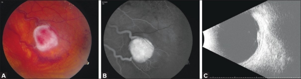
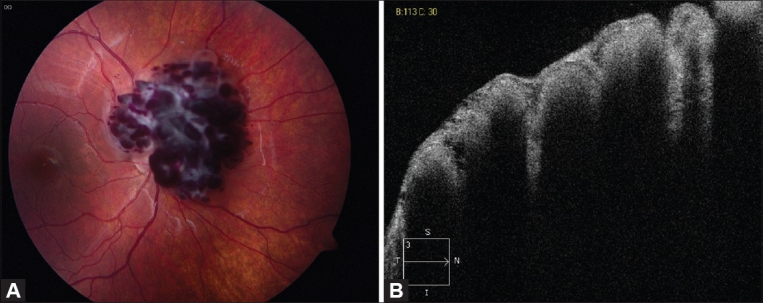
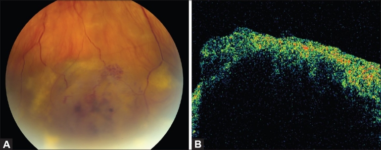
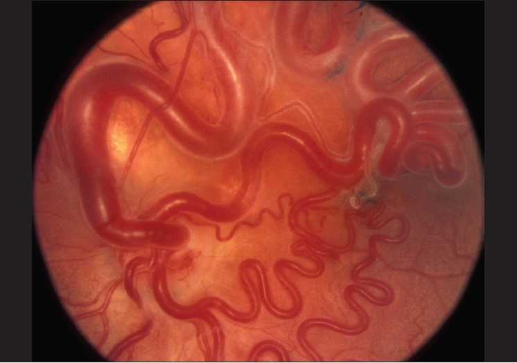
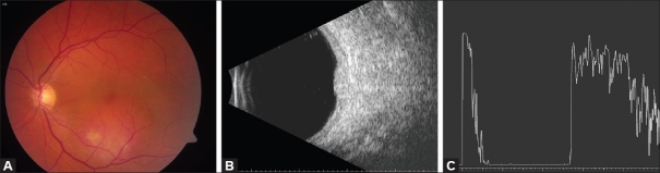
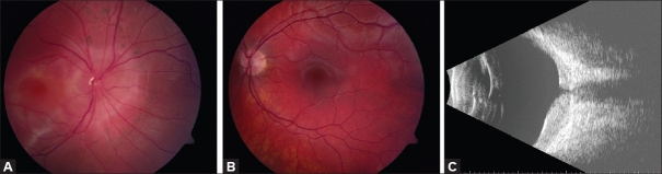
The vascular tumors of the retina and choroid comprise a diverse group of congenital and acquired lesions. The major vascular tumors of the retina include retinal capillary hemangioma, cavernous hemangioma of the retina, retinal vasoproliferative tumor, and racemose hemangiomatosis of the retina or Wyburn-Mason syndrome. Choroidal vascular tumors include circumscribed choroidal hemangioma and diffuse choroidal hemangioma. While classified as benign, visual symptoms secondary to exudative retinal detachment and a variety of other mechanisms are common and are a major source of long-term visual disability. While many therapeutic modalities exist, treatment of symptomatic cases can be challenging. Of particular importance, many of the vascular tumors of the retina and choroid have significant associations with systemic disease. As ocular symptoms are often the most common presenting disease manifestation, the ophthalmologist plays an important role in accurate and early diagnosis. The ability to initiate prompt screening and treatment in appropriate cases is critical. In the following article, the key clinical and diagnostic features of the major retinal and choroidal vascular tumors, their systemic associations, and the literature pertaining to the most currently available treatment strategies are reviewed.
Keywords: Cavernous Hemangioma; Choroidal Hemangioma; Retinal Capillary Hemangioma; Retinal Vasoproliferative Tumor; Wyburn–Mason Syndrome.
Conflict of interest statement
Figures
References
-
- Maher ER, Kaelin WG. Von Hippel-Lindau disease. Medicine. 1997;76:381–91. - PubMed
-
- Stolle C, Glenn G, Zbar B, Humphrey JS, Choyke P, Walther M, et al. Improved detection of germline mutation in the von Hippel-Lindau disease tumor suppression gene. Hum Mutat. 1998;12:417–23. - PubMed
-
- Chang JH, Spraul CW, Lynn ML, Drack A, Grossniklaus HE. The two-stage mutation model in retinal hemangioblastoma. Ophthalmic Genet. 1998;19:123–30. - PubMed
-
- Maher ER, Yates JR, Harries R, Benjamin C, Harris R, Moore AT, et al. Clinical features and natural history of von Hippel-Lindau disease. Q J Med. 1990;77:1151–63. - PubMed
-
- Richard S, Chauveau D, Chrétien Y, Beigelman C, Denys A, Fendler JP, et al. Renal lesions and pheochromocytomas in von Hippel-Lindau disease. Adv Nephrol Necker Hosp. 1994;23:1–27. - PubMed
LinkOut - more resources
Full Text Sources
Other Literature Sources
Research Materials
