

Small choroidal melanoma with monosomy 3
- PMID: 20844685
- PMCID: PMC2934721
- DOI: 10.4103/0974-9233.65487
Small choroidal melanoma with monosomy 3
Abstract
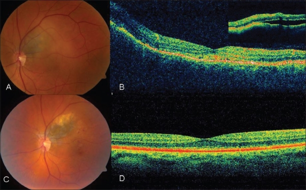
To report a patient with small juxtapapillary choroidal melanoma with chromosome 3 monosomy treated with I(125) plaque and transpupillary thermotherapy (TTT). A 64-year-old Caucasian male presented with painless blurred vision of the left eye. Ocular examination disclosed a small juxtapapillary choroidal melanocytic tumor with overlying subretinal fluid and orange pigment. Ultrasound showed an elevated choroidal mass of 2 mm thickness with low reflectivity on A-scan and hollowness on B scan, consistent with a small choroidal melanoma. The patient was treated with plaque I(125) radiotherapy combined with one session of TTT. Genetic testing of the tumor cells obtained by fine needle aspiration biopsy showed chromosome 3 monosomy. At 1 year after treatment, the tumor was regressed with resolution of subretinal fluid and 20/40 visual acuity. A small choroidal melanoma can manifest monosomy of chromosome 3, a known predictive factor for the development of systemic metastasis.
Keywords: Choroidal Melanoma; Chromosome 3; Eye; Metastases; Monosomy; Small Choroidal Melanoma.
Conflict of interest statement
Figures
Similar articles
-
Small choroidal melanoma with chromosome 3 monosomy on fine-needle aspiration biopsy.Ophthalmology. 2007 Oct;114(10):1919-24. doi: 10.1016/j.ophtha.2007.04.054. Epub 2007 Aug 15. Ophthalmology. 2007. PMID: 17698199
-
Primary transpupillary thermotherapy for choroidal melanoma in 391 cases: importance of risk factors in tumor control.Ophthalmology. 2015 Mar;122(3):600-9. doi: 10.1016/j.ophtha.2014.09.029. Epub 2014 Nov 13. Ophthalmology. 2015. PMID: 25439431
-
Primary transpupillary thermotherapy of choroidal melanocytic lesions.Middle East Afr J Ophthalmol. 2011 Apr;18(2):183-8. doi: 10.4103/0974-9233.80711. Middle East Afr J Ophthalmol. 2011. PMID: 21731333 Free PMC article.
-
[Choroidal melanoma: current therapeutic approaches].J Fr Ophtalmol. 2002 Feb;25(2):203-11. J Fr Ophtalmol. 2002. PMID: 11941244 Review. French.
-
Recent developments in the management of choroidal melanoma.Curr Opin Ophthalmol. 2004 Jun;15(3):244-51. doi: 10.1097/01.icu.0000120713.35941.e4. Curr Opin Ophthalmol. 2004. PMID: 15118513 Review.
References
-
- Shields CL, Shields JA, Kiratli H, De Potter P, Cater JR. Risk factors for growth and metastasis of small choroidal melanocytic lesions. Ophthalmology. 1995;102:1351–61. - PubMed
-
- Prescher G, Bornfeld N, Hirche H, Horsthemke B, Jöckel KH, Becher R. Prognostic implications of monosomy 3 in uveal melanoma. Lancet. 1996;347:1222–5. - PubMed
-
- Shields CL, Ganguly A, Materin MA, Teixeira L, Mashayekhi A, Swanson LA, et al. Chromosome 3 analysis of uveal melanoma using fine-needle aspiration biopsy at the time of plaque radiotherapy in 140 consecutive cases: The Deborah Iverson, MD, Lectureship. Arch Ophthalmol. 2007;125:1017–24. - PubMed
-
- Saida T. Lessons learned from studies of the development of early melanoma. Int J Clin Oncol. 2005;10:371–4. - PubMed
-
- Shields CL, Furuta M, Berman EL, Zahler JD, Hoberman DM, Dinh DH, et al. Choroidal nevus transformation into melanoma: Analysis of 2514 consecutive cases. Arch Ophthalmol. 2009;127:981–7. - PubMed
LinkOut - more resources
Full Text Sources
