

Review
The iris - a window into the genetics of common and rare eye diseases
Affiliations
- PMID: 20844723
- PMCID: PMC2938985
Item in Clipboard
Review
The iris - a window into the genetics of common and rare eye diseases
Ulster Med J.
2010 Jan.
Abstract
Visual examination, without instruments, of the eye allows inspection of the iris, sclera, cornea and, through the iris, some abnormalities of the lens and retina. Several hereditary disorders can easily be recognised by characteristic iris changes. This review discusses changes in the iris, visible lens anomalies, and changes in the cornea surrounding the iris. A genetic diagnosis can help with management of diseases. Some conditions are single gene disorders, some are chromosomal rearrangements, and some are abnormalities of fetal development.
Figures

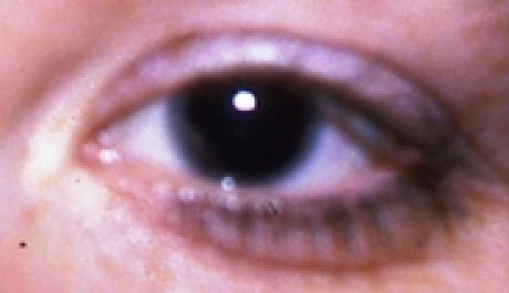
NHS evidence ‘normal’ iris.

From left to right, abnormal irides – see text for diagnoses.

From left to right, abnormal irides – see text for diagnoses.

From left to right, abnormal irides – see text for diagnoses.

From left to right, abnormal irides – see text for diagnoses.

From left to right, abnormal irides – see text for diagnoses.

From left to right, abnormal irides – see text for diagnoses.

From left to right, abnormal irides – see text for diagnoses.

From left to right, abnormal irides – see text for diagnoses.

From left to right, abnormal irides – see text for diagnoses.
From left to right, abnormal irides – see text for diagnoses.

From left to right, abnormal irides – see text for diagnoses.

From left to right, abnormal irides – see text for diagnoses.

From left to right, abnormal irides – see text for diagnoses.

From left to right, abnormal irides – see text for diagnoses.

From left to right, abnormal irides – see text for diagnoses.

From left to right, abnormal irides – see text for diagnoses.

From left to right, abnormal irides – see text for diagnoses.

From left to right, abnormal irides – see text for diagnoses.

X-linked cataract.

Epibulbar dermoid overlying the iris.

Rieger anomaly.

Autosomal dominant family tree. Affected cases are shaded.
Publication types
MeSH terms
LinkOut - more resources
Full Text Sources