. 2010 Sep 10;5(9):e12599.
doi: 10.1371/journal.pone.0012599.

The CC-NB-LRR-type Rdg2a resistance gene confers immunity to the seed-borne barley leaf stripe pathogen in the absence of hypersensitive cell death

Affiliations

The CC-NB-LRR-type Rdg2a resistance gene confers immunity to the seed-borne barley leaf stripe pathogen in the absence of hypersensitive cell death

Davide Bulgarelli et al. PLoS One. .

Abstract

Background: Leaf stripe disease on barley (Hordeum vulgare) is caused by the seed-transmitted hemi-biotrophic fungus Pyrenophora graminea. Race-specific resistance to leaf stripe is controlled by two known Rdg (Resistance to Drechslera graminea) genes: the H. spontaneum-derived Rdg1a and Rdg2a, identified in H. vulgare. The aim of the present work was to isolate the Rdg2a leaf stripe resistance gene, to characterize the Rdg2a locus organization and evolution and to elucidate the histological bases of Rdg2a-based leaf stripe resistance.

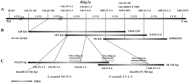
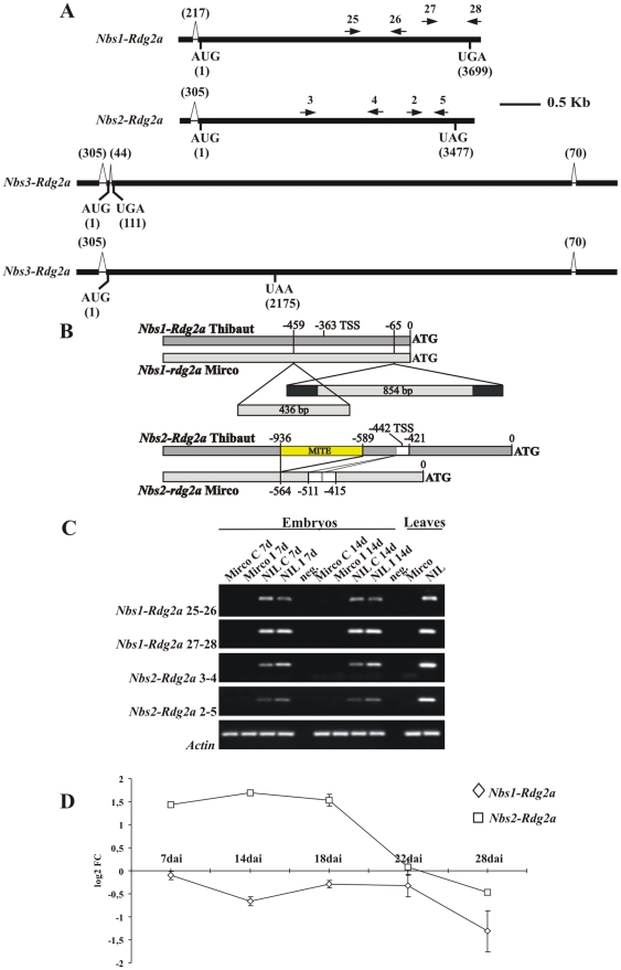
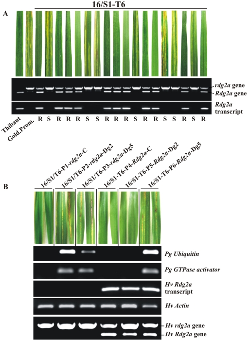
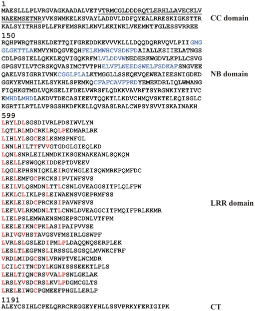
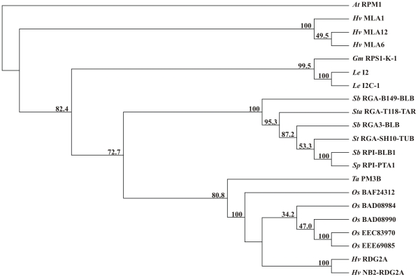
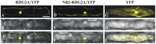
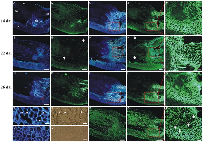
Principal findings: We describe here the positional cloning and functional characterization of the leaf stripe resistance gene Rdg2a. At the Rdg2a locus, three sequence-related coiled-coil, nucleotide-binding site, and leucine-rich repeat (CC-NB-LRR) encoding genes were identified. Sequence comparisons suggested that paralogs of this resistance locus evolved through recent gene duplication, and were subjected to frequent sequence exchange. Transformation of the leaf stripe susceptible cv. Golden Promise with two Rdg2a-candidates under the control of their native 5' regulatory sequences identified a member of the CC-NB-LRR gene family that conferred resistance against the Dg2 leaf stripe isolate, against which the Rdg2a-gene is effective. Histological analysis demonstrated that Rdg2a-mediated leaf stripe resistance involves autofluorescing cells and prevents pathogen colonization in the embryos without any detectable hypersensitive cell death response, supporting a cell wall reinforcement-based resistance mechanism.

Conclusions: This work reports about the cloning of a resistance gene effective against a seed borne disease. We observed that Rdg2a was subjected to diversifying selection which is consistent with a model in which the R gene co-evolves with a pathogen effector(s) gene. We propose that inducible responses giving rise to physical and chemical barriers to infection in the cell walls and intercellular spaces of the barley embryo tissues represent mechanisms by which the CC-NB-LRR-encoding Rdg2a gene mediates resistance to leaf stripe in the absence of hypersensitive cell death.


Conflict of interest statement

Competing Interests: The authors have declared that no competing interests exist.

Figures

Figure 1
Figure 1. Genetic and physical maps of the Rdg2a locus.
(A) Genetic map of Rdg2a. Crossovers identified in the 1,400 F2 plants from a cross between Thibaut (Rdg2a) and Mirco are shown at the top (CO). Orientation is indicated by Tel (telomere) and Cen (centromere). (B) Contig of Morex BAC clones. (C) Thibaut cosmid contig and genes at the Rdg2a locus. Transcription direction of the genes are indicated by arrows.
Figure 2
Figure 2. Analysis of Rdg2a-candidate gene transcript structure and regulation.
(A) Nbs1-Rdg2a, Nbs2-Rdg2a and Nbs3-Rdg2a transcript structures (cv. Thibaut), indicating positions of primers used in transcript quantification. The two transcript types resulting from alternative splicing pattern of Nbs3-Rdg2a are indicated. (B) Structural differences between Thibaut and Mirco alleles of Nbs1-rdg2a and Nbs2-rdg2a genes in 5′ regions. Positions of insertion/deletions relative to the start codon are shown. Filled sections indicate inverted repeats present in an insertion in the Mirco Nbs1-rdg2a gene. The Nbs2-rdg2a allele comparison illustrates variation for a MITE insertion and a 41–bp direct repeat (open sections). Transcription start sites (TSS) for Nbs1-Rdg2a and Nbs2-Rdg2a are indicated. (C) Semi-quantitative RT-PCR analysis of the Rdg2a-candidate gene expression using gene specific primers. Transcripts were analysed in embryos of the cv. Mirco (rdg2a) and NIL3876 (Rdg2a) genotypes at two timepoints, after inoculation with P. graminea Dg2 (I), or in uninoculated controls (C). Leaves of uninoculated plants were also analysed. Negative controls (neg.) in which DNA was omitted are included. Primers for cv. Thibaut genes were those represented in (A), while primers for amplifying homologous fragments from cv. Mirco were based on the cv. Mirco gene sequences and positioned within 30 bp of the corresponding Thibaut primers. RT-PCR of the barley β-actin gene was used as an internal control. (D) Quantitative RT-PCR at 7, 14, 18, 22 and 28 days after pathogen inoculation (dai) for the two Rdg2a-candidates in embryos of NIL3876-Rdg2a. Values are expressed as log2 fold changes of transcript levels in the inoculated samples with respect to the transcript levels in un-inoculated barley embryos. Error bars represent SD across all RT-PCR replicates (four to six from each of two independent inoculations).
Figure 3
Figure 3. Analysis of T1 family 16/S1-T6 segregating for the Nbs1-Rdg2a transgene.
(A) T1 seeds were inoculated with P. graminea isolate Dg2 and plants analyzed for disease symptoms in leaves (upper panel), an STS marker for Rdg2a (middle panel; upper band represents the rdg2a susceptibility allele from cv. Golden Promise while the lower band represents the Rdg2a transgene or endogenous gene), and Rdg2a transgene or endogenous gene expression by RT-PCR (lower panel). Resistance (R) or susceptibility (S) status of the plants is indicated underneath. The resistant cv. Thibaut and the susceptible cv. Golden Promise provide controls. (B) Leaves of six 16/S1-T6 T1 plants were analysed for expression of the fungal (Pg) Ubiquitin and GTPase activator genes and the barley (Hv) Rdg2a gene by RT-PCR. Seeds had been inoculated with Dg2 or Dg5 leaf stripe isolates or were non-inoculated (C). The barley β-actin gene was used as an internal control. Plant DNA was also tested for the presence of the transgene using the Rdg2a STS marker described in (A).
Figure 4
Figure 4. RDG2A protein sequence.
The predicted coiled-coil (CC) domain is underlined. Motifs conserved in the NB region of NB-LRR proteins are in blue, and are (in order): P-loop, RNBS-A, Kinase 2, RNBS-C, GLPL, RNBS-D and MHD. Amino acids conforming to the cytoplasmic LRR consensus LxxLxLxx(C/N/T)xxLxxLxxLP are in red. CT denotes the RDG2A C-terminal region.
Figure 5
Figure 5. Neighbor-joining phylogenetic tree including RDG2A and similar resistance proteins and resistance gene analog products.
Numbers on the branches indicate bootstrap percentages. Prefixes indicate species origin. The A. thaliana RPM1 protein (Q39214) was used as an outgroup. Shown are the rice (Oryza sativa) disease resistance-like proteins BAF24312, BAD08984, BAD08990, EEC83970 and EEE69085, the PM3 wheat powdery mildew resistance protein, products of the S. bulbocastaneum blight resistance gene Rpi-blb1 and its paralogues Rga3-blb, and Rpi-blb1, predicted products of RGA_B149.blb, RGA_T118-tar (S. tarijense), RGA_SH10-tub (S. tuberosum) and Rpi-pta1 (S. papita), the I2 and I2C-1 proteins encoded by the tomato (Lycopersicon esculentum) I2 resistance locus to Fusarium wilt, the soybean (Glycine max) Phytophthora root rot resistance protein RPS-L-K-1, and the barley (H. vulgare) powdery mildew resistance proteins MLA1, MLA6 and MLA12.
Figure 6
Figure 6. Sub-cellular localization of RDG2A and NB2-RDG2A proteins.
Barley cv. Golden Promise epidermal cells were transiently transformed with constructs expressing RDG2A:YFP and NB2-RDG2A:YFP fusion proteins (A and D respectively), driven by the maize polyubiquitin gene promoter. A construct expressing YFP alone with the same promoter was used as control (G). Fluorescence signals were visualized using confocal laser scanning microscopy (A, D and G). Bright field images (B, E and H) and merged images (C, F and I) are shown. Scale bar represent 50 µm.
Figure 7
Figure 7. Histological analyses of NIL3876-Rdg2a barley embryos.
(A) to (C) Sections of embryos grown under control conditions observed under UV excitation. (D) to (F) Sections in (A) to (C) subjected to TUNEL analysis. (G) to (I) Sections of embryos inoculated with leaf stripe isolate Dg2 and observed under UV excitation. (J) to (L) Sections in (G) to (I) subjected to TUNEL analysis; the bright green fluorescence at the level of scutellar node and provascular tissue is due to cell wall autofluorescence. (M) to (O) Magnified views of the boxed regions in (J) to (L) and (G) to (I). (S) and (T) Magnified views of the smaller box in (I) stained with calcofluor (S) or observed under bright field (T); arrows indicate the intercellularly growing P. graminea mycelium. (U) and (V) Magnified views of the small box in (C) stained with calcofluor (U) or observed under bright field (V). (P) and (Q) Respectively, sections of control and inoculated embryos at 26 dai, treated with DNase I and subjected to TUNEL analysis. (R) A magnified view of the region boxed in (Q). White arrows in Figure 7E, K and R indicate TUNEL positive nuclei. Scale bars represent 200 µM (A) to (L), 50 µM (M) to (O) and 25 µM (S) to (T). co  =  coleoptile, pt  =  provascular tissue, sa  =  shoot apex, sn  =  scutellar node.

Similar articles

Cited by

References

    1. Delogu G, Porta-Puglia A, Stanca AM, Vannacci G. Interaction between barley and Pyrenophora graminea: an overview of research in Italy. Rachis. 1995;14:29–34.
    1. Mueller KJ, Valè G, Enneking D. Selection of resistant spring barley accessions after natural infection with leaf stripe (Pyrenophora graminea) under organic farming conditions in Germany and by sandwich test. J Plant Pathol. 2003;85:9–14.
    1. Platenkamp R. Investigations on the infections pathway of Drechslera graminea in germinating barley. Royal Veterinary and Agricultural University, Yearbook. 1976:49–64.
    1. Hammouda AM. Variability of Drechslera graminea, the causal fungus of leaf stripe of barley. Acta Phytopathol Entomol Hung. 1988;23:73–80.
    1. Haegi A, Bonardi V, Dall'Aglio E, Glissant D, Tumino G, et al. Histological and molecular analysis of Rdg2a barley resistance to leaf stripe. Mol Plant Pathol. 2008;9:463–478. - PMC - PubMed

Publication types

MeSH terms