. 2011 Jan-Feb;17(1-2):12-20.
doi: 10.2119/molmed.2010.00103. Epub 2010 Sep 10.

High expression of H3K27me3 in human hepatocellular carcinomas correlates closely with vascular invasion and predicts worse prognosis in patients

Affiliations

High expression of H3K27me3 in human hepatocellular carcinomas correlates closely with vascular invasion and predicts worse prognosis in patients

Mu-Yan Cai et al. Mol Med. 2011 Jan-Feb.

Abstract

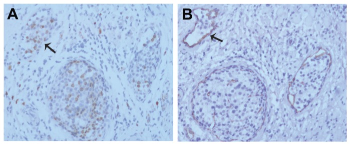
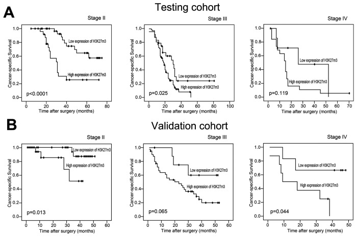
It has been suggested that trimethylation of lysine 27 on histone H3 (H3K27me3) is a crucial epigenetic process in tumorigenesis. However, the expression dynamics of H3K27me3 and its clinicopathological/prognostic significance in hepatocellular carcinoma (HCC) are unclear. In this study, immunohistochemical analysis (IHC) was used to examine protein expression of H3K27me3 in HCC tissues from two independent cohorts and corresponding nontumorous hepatocellular tissues by tissue microarray. The optimal cutpoint of H3K27me3 expression was assessed by the X-tile program. Our results showed that the cutpoint for high expression of H3K27me3 in HCCs was determined when more than 70% of the tumor cells showed positive staining. High expression of H3K27me3 was observed in 134 of 212 (63.2%) and 76 of 126 (60.4%) of HCCs in the testing and validation cohorts, respectively. Correlation analysis demonstrated that high expression of H3K27me3 in HCCs was significantly correlated with large tumor size, multiplicity, poor differentiation, advanced clinical stage and vascular invasion (P < 0.05). In addition, high expression of H3K27me3 in HCC patients was associated closely with shortened survival time, independent of serum α-fetoprotein levels, tumor size and multiplicity, clinical stage, vascular invasion and relapse as evidenced by univariate and multivariate analysis in both cohorts (P < 0.05). In different subsets of HCC patients, H3K27me3 expression was also a prognostic indicator in patients with stage II tumors (P < 0.05). Thus, these findings provide evidence that a high expression of H3K27me3, as detected by IHC, correlates closely with vascular invasion of HCCs and is an independent molecular marker for poor prognosis in patients with HCC.


Figures

Figure 1
Figure 1
The expression of H3K27me3 in HCC and adjacent nonneoplastic liver tissues by IHC. (A) High expression of H3K27me3 was shown in an HCC patient sample (case 68 in the testing cohort), in which more than 95% carcinoma cells revealed positive staining of H3K27me3 in nuclei (100×). (B) Another HCC case in the testing cohort (case 36) demonstrated low expression of H3K27me3, in which less than 30% of tumor cells showed immunoreactivity of H3K27me3 in nuclei (100×). (C) Negative expression of H3K27me3 was detected in an HCC case (case 70) in the testing cohort (100×). (D) Adjacent non-neoplastic liver tissues of the same HCC case 36 showed nearly negative expression of H3K27me3 protein, in which less than 2% of normal hepatic cells showed positive staining of H3K27me3 in nuclei (100×). (E), (F), (G) and (H) demonstrate the higher magnification (400×) from the area of the box in (A), (B), (C) and (D), respectively.
Figure 2
Figure 2
X-tile plots of the prognostic marker H3K27me3 on HCC cohorts. X-tile analysis was carried out on patient data from the testing cohort, equally divided into training and validation subsets. X-tile plots of training sets are displayed in the left panels, with matched validation sets in the smaller inset. The plot showed the χ2 log-rank values created when the cohort was divided into two populations. The cutpoint highlighted by the black/white circle in the left panels was demonstrated on a histogram of the entire cohort (middle panels) and a Kaplan–Meier plot (right panels). P values were defined by using the cutpoint derived from a training subset to parse a separate validation subset. (A) H3K27me3 expression was divided at the optimal cutpoint, as defined by the most significant on the plot (≤70% and >70% of tumor cells with positive staining of H3K27me3; P < 0.0001). (B) The optimal cutpoints for H3K27me3 expression determined by X-tile plot of the testing cohort were applied to the validation cohort and reached high statistical significance (P < 0.0001).
Figure 3
Figure 3
High expression of H3K27me3 was associated with HCC vascular invasion. (A) Higher expression of H3K27me3 was observed in carcinoma cells located at blood vessels in a patient with HCC (case 89 in testing cohort, arrow, 200×). (B) The endothelial cells of blood vessels (arrow) were stained for IHC, with an anti-CD34 antibody on the serial section of the same case 89 (200×).
Figure 4
Figure 4
Kaplan–Meier survival analysis of H3K27me3 expression in subsets of HCC patients in different stages (log-rank test). (A) Stage II, probability of survival of stage II patients with HCC in the testing cohort: low expression, n = 33; high expression, n = 27. Stage III, probability of survival of patients with stage III HCC in the testing cohort: low expression, n = 24; high expression, n = 73. Stage IV, probability of survival of stage IV patients with HCC in the testing cohort: low expression, n = 8; high expression, n = 19. (B) Stage II, probability of survival of stage II patients with HCC in the validation cohort: low expression, n = 29; high expression, n = 21. Stage III, probability of survival of stage III patients with HCC in the validation cohort: low expression, n = 9; high expression, n = 41. Stage IV, probability of survival of stage IV patients with HCC in the validation cohort: low expression, n = 6; high expression, n = 8.
Figure 5
Figure 5
ROC curve analysis for different clinicopathological features and H3K27me3 expression was performed to evaluate the survival status. (A) Sex (AUC = 0.539; P = 0.339), age (AUC = 0.504; P = 0.923), serum AFP (AUC = 0.678; P < 0.0001), hepatitis history (AUC = 0.510; P = 0.812), cirrhosis (AUC = 0.503; P = 0.942), tumor multiplicity (AUC = 0.687; P < 0.0001), tumor size (AUC = 0.699; P < 0.0001), differentiation (AUC = 0.597; P = 0.017) stage (AUC = 0.746; P < 0.0001), H3K27me3 expression (AUC = 0.733; P < 0.0001), vascular invasion (AUC = 0.764; P < 0.0001), and relapse (AUC = 0.664; P < 0.0001) implied statistical associations with survival in the testing cohort. (B) Sex (AUC = 0.508; P = 0.880), age (AUC = 0.521; P = 0.695), serum AFP (AUC = 0.702; P < 0.0001), hepatitis history (AUC = 0.504; P = 0.940), cirrhosis (AUC = 0.491; P = 0.868), tumor multiplicity (AUC = 0.710; P < 0.0001), tumor size (AUC = 0.715; P = 0.002), differentiation (AUC = 0.612; P = 0.036) stage (AUC = 0.761; P < 0.0001), H3K27me3 expression (AUC = 0.719; P < 0.0001), vascular invasion (AUC = 0.757; P < 0.0001) and relapse (AUC = 0.712; P < 0.0001) were used to test the survival status in validation cohort.

References

    1. Ince N, Wands JR. The increasing incidence of hepatocellular carcinoma. N Engl J Med. 1999;340:798–9. - PubMed
    1. El-Serag HB, Mason AC. Rising incidence of hepatocellular carcinoma in the United States. N Engl J Med. 1999;340:745–50. - PubMed
    1. Hsu YC, Fu HH, Jeng YM, Lee PH, Yang SD. Proline-directed protein kinase FA is a powerful and independent prognostic predictor for progression and patient survival of hepatocellular carcinoma. J Clin Oncol. 2006;24:3780–8. - PubMed
    1. Nagao T, et al. Postoperative recurrence of hepatocellular carcinoma. Ann Surg. 1990;211:28–33. - PMC - PubMed
    1. Mann CD, et al. Prognostic molecular markers in hepatocellular carcinoma: a systematic review. Eur J Cancer. 2007;43:979–92. - PubMed

Publication types

MeSH terms