. 2011 Feb;469(2):429-36.
doi: 10.1007/s11999-010-1554-7.

Hip offset in total hip arthroplasty: quantitative measurement with navigation

Affiliations

Hip offset in total hip arthroplasty: quantitative measurement with navigation

Manish Dastane et al. Clin Orthop Relat Res. 2011 Feb.

Abstract

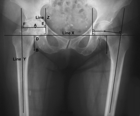
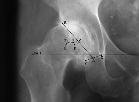
Background: Offset in THA correlates to abductor muscle function, wear, and impingement. Femoral offset after THA is not independent of the cup center of rotation (COR) so hip offset, a combination of femoral offset and change in hip COR, becomes the important measurement.

Questions/purposes: We therefore asked whether hip offset in arthritic hips would correlate with cup COR; whether offset could always be balanced within 6 mm of contralateral normal hips; and whether hip length could also be kept within 6 mm.

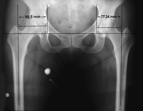
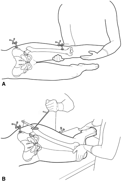
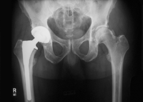
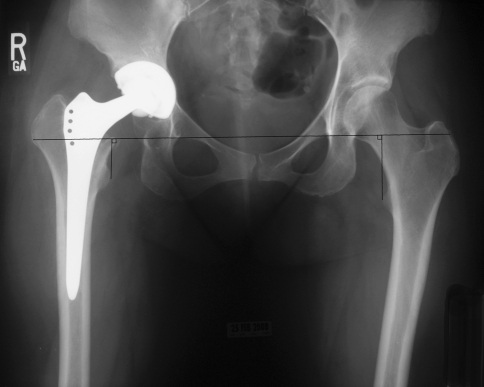
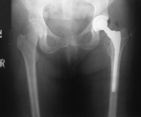
Methods: We compared hip offset of arthritic and contralateral normal hips on radiographs in 82 patients (82 hips) who had THA. We used computer navigation in all patients with the aim of reconstructing the hip offset and to compare hip offset change to the quantitative change of the hip COR.

Results: The preoperative radiographic change to equalize the offset ranged from -12 to +21 mm (mean, 1.5); postoperatively the change was 1.4 ± 6.4 mm and was within ± 6 mm in 78 of 82 hips. As COR displaced superiorly from 3 to 6+ mm the offset had to be substantially increased. Only with COR 0-3 mm superior and 0-5 mm medial was offset always within 5 mm.

Conclusions: Hip offset reconstruction was directly related to the position of the hip COR, and navigation allowed quantitative control of offset and hip length.


Figures

Fig. 1
Fig. 1
The method for measuring hip length and hip offset is shown. Point A is the center of the femoral head, point B is the apex of the lesser trochanter, and line Y is the anatomic axis of the femur. Hip offset is measured as follows: Line X is the transteardrop line with line Z drawn through the teardrop perpendicular to the transteardrop line. A perpendicular line between line Y and line Z through the femoral head hip center (point A) is the hip offset, ie, AE plus AF. Hip length is measured from the apex of the lesser trochanter, point B, perpendicular to the transteardrop line X, line BD.
Fig. 2
Fig. 2
Preoperative AP pelvis radiograph shows hips with asymmetrical offset. The arthritic hip has femoral head migration of 5.56 mm in the horizontal direction which means the radiographic anatomic asymmetry is 16.7 mm.
Fig. 3
Fig. 3
Line T designates the transteardrop line. Line XY designates the transverse acetabular ligament. The mouth of acetabulum is from the midpoint of Line XY (point A) to the superior-lateral cortical rim of the acetabulum excluding osteophytes (point B). Point Z marks the midpoint of Line AB. Line ZD is 7 mm lateral and perpendicular to point Z. The center of the acetabulum (point C) is 1 mm cranial to point D. The center of the femoral head (F) was determined by using a transparent template with concentric circles. Line CE is drawn parallel to the transteardrop line from point C to point F. The change necessary to make the centers of rotation C and F coincide in the lateral direction (offset) is Line CE and the vertical direction (hip length) is Line FE.
Fig. 4A–B
Fig. 4A–B
(A) A diagram shows the use of the femoral tracker array for leg length and offset. Leg position is monitored by the software to be exactly duplicated after reconstruction. (B) A diagram shows femur registration using a screw in the greater trochanter. The pointer touches the depression of the screw head for the measurement before and after reconstruction. The same surgical assistant positions the legs by aligning the patellae and heels of the feet.
Fig. 5
Fig. 5
Postoperative radiographic with hip COR within 3 mm superior and 5 mm medial so hip offset and length are within 3 mm of the normal hip.
Fig. 6
Fig. 6
Postoperative AP pelvis radiograph of dysplastic hip with acetabular cup placement using the medial protrusio technique which elevated and medialized the COR more than 6 mm so the reconstruction required a high offset stem. This was the only hip which did not keep hip length discrepancy within 6 mm.
Fig. 7
Fig. 7
Postoperative radiograph with offset increased more than 6 mm to prevent bony impingement because of a valgus hip. The iliopsoas tendon should be recessed to avoid tendinitis.

References

    1. Asayama I, Chamnongkich S, Simpson KJ, Kinsey TL, Mahoney OM. Reconstructed hip joint position and abductor muscle strength after total hip arthroplasty. J Arthroplasty. 2005;20:414–420. doi: 10.1016/j.arth.2004.01.016. - DOI - PubMed
    1. Bourne RB, Rorabeck CH. Soft tissue balancing: The hip. J Arthroplasty. 2002;17(Suppl 1):17–22. doi: 10.1054/arth.2002.33263. - DOI - PubMed
    1. Charles MN, Bourne RB, Davey JR, Greenwald AS, Morrey BF, Rorabeck CH. Soft-tissue balancing of the hip: the role of femoral offset restoration. J Bone Joint Surg Am. 2004;86:1078–1088. - PubMed
    1. Dolhain P, Tsigaras H, Bourne RB, Rorabeck CH, Mac Donald S, Mc Calden R. The effectiveness of dual offset stems in restoring offset during total hip replacement. Acta Orthop Belg. 2002;68:490–499. - PubMed
    1. Dorr LD. Hip Arthroplasty: Minimally Invasive Techniques and Computer Navigation. 1. Philadelphia, PA: WB Saunders; 2005.

MeSH terms