

Current approaches to measuring human islet-antigen specific T cell function in type 1 diabetes
- PMID: 20846160
- PMCID: PMC2996587
- DOI: 10.1111/j.1365-2249.2010.04237.x
Current approaches to measuring human islet-antigen specific T cell function in type 1 diabetes
Abstract
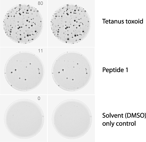
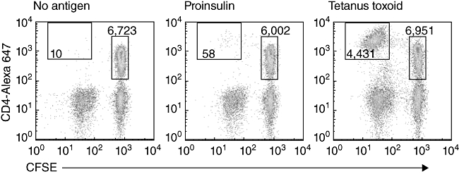
Type 1 diabetes (T1D) is an autoimmune disease caused by the T cell-mediated destruction of the pancreatic insulin-producing beta cells. Currently there are no widely accepted and standardized assays available to analyse the function of autoreactive T cells involved in T1D. The development of such an assay would greatly aid efforts to understand the pathogenesis of T1D and is also urgently required to guide the development of antigen-based therapies intended to prevent, or cure, T1D. Here we describe some of the assays used currently to detect autoreactive T cells in human blood and review critically their strengths and weaknesses. The challenges and future prospects for the T cell assays are discussed.
© 2010 The Authors. Clinical and Experimental Immunology © 2010 British Society for Immunology.
Figures
References
-
- Atkinson MA, Maclaren NK. The pathogenesis of insulin-dependent diabetes mellitus. N Engl J Med. 1994;331:1428–36. - PubMed
-
- Santamaria P, Utsugi T, Park BJ, Averill N, Kawazu S, Yoon JW. Beta-cell-cytotoxic CD8+ T cells from nonobese diabetic mice use highly homologous T cell receptor alpha-chain CDR3 sequences. J Immunol. 1995;154:2494–503. - PubMed
-
- Keymeulen B, Vandemeulebroucke E, Ziegler AG, et al. Insulin needs after CD3-antibody therapy in new-onset type 1 diabetes. N Engl J Med. 2005;352:2598–608. - PubMed
Publication types
MeSH terms
Substances
LinkOut - more resources
Full Text Sources
Other Literature Sources
Medical
