. 2010 Dec;162(3):474-86.
doi: 10.1111/j.1365-2249.2010.04256.x. Epub 2010 Sep 15.

Inflammatory conditions affect gene expression and function of human adipose tissue-derived mesenchymal stem cells

Affiliations

Inflammatory conditions affect gene expression and function of human adipose tissue-derived mesenchymal stem cells

M J Crop et al. Clin Exp Immunol. 2010 Dec.

Abstract

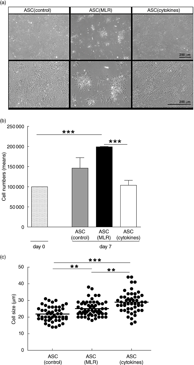
There is emerging interest in the application of mesenchymal stem cells (MSC) for the prevention and treatment of autoimmune diseases, graft-versus-host disease and allograft rejection. It is, however, unknown how inflammatory conditions affect phenotype and function of MSC. Adipose tissue-derived mesenchymal stem cells (ASC) were cultured with alloactivated peripheral blood mononuclear cells (PBMC) (mixed lymphocyte reaction: MLR), with proinflammatory cytokines [interferon (IFN)-γ, tumour necrosis factor (TNF)-α and interleukin (IL)-6] or under control conditions, and their full genome expression and function examined. Proinflammatory cytokines mainly increased indoleamine-2,3-dioxygenase expression, whereas ASC cultured with MLR showed increased expression of COX-2, involved in prostaglandin E(2) production. Both conditions had a stimulatory, but differential, effect on the expression of proinflammatory cytokines and chemokines, while the expression of fibrotic factors was decreased only in response to proinflammatory cytokines. Functional analysis demonstrated that inflammatory conditions affected morphology and proliferation of ASC, while their differentiation capacity and production of trophic factors was unaffected. The immunosuppressive capacity of ASC was enhanced strongly under inflammatory conditions. In conclusion, ASC showed enhanced immunosuppressive capacity under inflammatory conditions, while their differentiation capacity was preserved. Therefore, in vitro preconditioning provides ASC with improved properties for immediate clinical immune therapy.


Figures

Fig. 1
Fig. 1
Hierarchical clustering of genes up- (red) and down-regulated (green) by adipose tissue-derived mesenchymal stem cells (ASC) after culture under inflammatory conditions compared to average gene expression level of all samples, measured by microarray. ASC were cultured under control conditions, with alloactivated peripheral blood mononuclear cells (PBMC) (mixed lymphocyte reaction: MLR) or with proinflammatory cytokines (n = 4). Microarray data is deposited in Gene Expression Omnibus (GEO) number GSE18662 at http://www.ncbi.nlm.nih.gov/geo/query/acc.cgi?acc=GSE18662. (a) Gene expression profile of ASC cultured with MLR compared to control ASC. ASC cultured with MLR showed partial overlap with ASC cultured with proinflammatory cytokines. (b) Gene expression profile of ASC cultured with proinflammatory cytokines compared to control ASC. ASC cultured with proinflammatory cytokines showed partial overlap with ASC cultured with MLR. (c) Gene expression profile of ASC cultured with MLR compared to ASC cultured with proinflammatory cytokines. ASC cultured with inflammatory conditions showed partial overlap with control ASC.
Fig. 2
Fig. 2
Gene expression of adipose tissue-derived mesenchymal stem cells (ASC) cultured under control conditions, with alloactivated peripheral blood mononuclear cells (PBMC) (mixed lymphocyte reaction: MLR) or with proinflammatory cytokines for 7 days. mRNA expression is shown as log2-transformed median fluorescence intensities of four independent samples as measured by microarray. (a) Immunosuppressive factors; (b) guanylate binding proteins; (c) myxovirus resistance genes; (d) proinflammatory factors; (e) serum amyloid; (f) human leucocyte antigen (HLA) class I; (g) HLA class II; (h) chemokine ligands (CXCL); (i) chemokine ligands (CCL); (j) collagens.
Fig. 3
Fig. 3
Effect of inflammatory conditions on the phenotype of adipose tissue-derived mesenchymal stem cells (ASC). ASC were cultured under control conditions, with alloactivated peripheral blood mononuclear cells (PBMC) (mixed lymphocyte reaction: MLR) in transwell culture systems or with proinflammatory cytokines. For each independent experiment, ASC were used from different ASC donors. (a) Morphology of ASC. ASC cultured under control conditions and with proinflammatory cytokines formed confluent monolayers, while ASC co-cultured with MLR formed star-like clusters of cells. Representative examples shown of four experiments with four different ASC cultures. (b) Proliferation of ASC. Absolute number of viable ASC was counted after 7 days in culture. Data represent the mean ± standard error of the mean of six experiments with six different ASC cultures. (c) Cell size of ASC. ASC cultured with MLR and proinflammatory cytokines increased significantly in size compared to ASC cultured under control conditions. Approximately 50 individual trypsinized ASC per culture condition were measured. (d) Flow cytometric analysis of ASC. One representative example of each condition is shown of three independent experiments using three different ASC cultures. *P < 0·05; **P < 0·01; ***P < 0·001.
Fig. 3
Fig. 3
Effect of inflammatory conditions on the phenotype of adipose tissue-derived mesenchymal stem cells (ASC). ASC were cultured under control conditions, with alloactivated peripheral blood mononuclear cells (PBMC) (mixed lymphocyte reaction: MLR) in transwell culture systems or with proinflammatory cytokines. For each independent experiment, ASC were used from different ASC donors. (a) Morphology of ASC. ASC cultured under control conditions and with proinflammatory cytokines formed confluent monolayers, while ASC co-cultured with MLR formed star-like clusters of cells. Representative examples shown of four experiments with four different ASC cultures. (b) Proliferation of ASC. Absolute number of viable ASC was counted after 7 days in culture. Data represent the mean ± standard error of the mean of six experiments with six different ASC cultures. (c) Cell size of ASC. ASC cultured with MLR and proinflammatory cytokines increased significantly in size compared to ASC cultured under control conditions. Approximately 50 individual trypsinized ASC per culture condition were measured. (d) Flow cytometric analysis of ASC. One representative example of each condition is shown of three independent experiments using three different ASC cultures. *P < 0·05; **P < 0·01; ***P < 0·001.
Fig. 4
Fig. 4
Effect of inflammatory conditions on the differentiation capacity of adipose tissue-derived mesenchymal stem cells (ASC). ASC cultured under control conditions, with mixed lymphocyte reaction (MLR) or with proinflammatory cytokines, were continued in culture for 3 weeks under osteogenic or adipogenic conditions. One representative example is shown of four experiments using four different ASC donors. (a) Osteogenic differentiation. Deposition of calcified nodules (brown/black) was visualized by von Kossa staining. (b) Adipogenic differentiation. Lipid droplets (red) were stained with oil-red-O.
Fig. 5
Fig. 5
Effect of inflammatory conditions on the inhibition of peripheral blood mononuclear cell (PBMC) proliferation by adipose tissue-derived mesenchymal stem cells (ASC). ASC were pretreated under control conditions, with alloactivated PBMC mixed lymphocyte reaction (MLR) or with proinflammatory cytokines conditions and added to mitogen- or alloactivated PBMC. The proliferation in the test conditions is normalized to the proliferation in the control situation (i.e. MLR without addition of ASC, which is set to 100%). (a) Effect of pretreated ASC on the proliferation of phytohaemagglutinin (PHA)-stimulated PBMC. ASC were added to PBMC at various concentrations (1:5, 1:20, 1:50) (n = 8). Proliferation was measured by [3H]-thymidine incorporation on day 3. (b) Effect of pretreated ASC added on day 0 to MLR on the proliferation of alloactivated PBMC (MLR). ASC were added to PBMC at various concentrations (1:5, 1:20, 1:50) (n = 6). Proliferation was measured by [3H]-thymidine incorporation on day 7. (c) Effect of pretreated ASC added on day 6 to MLR. ASC were added to responder PBMC at a 1:5 ratio (n = 11). Proliferation was measured by [3H]-thymidine incorporation on day 7. (d) Effect of IDO1-inhibitor 1-methyl-l-tryptophan (1-MT) on the capacity of pretreated ASC to inhibit the proliferation of PHA-stimulated PBMC (n = 4). ASC were added to responder PBMC at a 1:5 ratio. Proliferation was measured by [3H]-thymidine incorporation on day 3. (e) Effect of IDO1-inhibitor 1-MT on the capacity of pretreated ASC to inhibit the proliferation in MLR (n = 8). ASC were added to responder PBMC at a 1:5 ratio. Proliferation was measured by [3H]-thymidine incorporation on day 7. *P < 0·05; **P < 0·01; ***P < 0·001.

References

    1. Toma JG, McKenzie IA, Bagli D, Miller FD. Isolation and characterization of multipotent skin-derived precursors from human skin. Stem Cells. 2005;23:727–37. - PubMed
    1. Pittenger MF, Mackay AM, Beck SC, et al. Multilineage potential of adult human mesenchymal stem cells. Science. 1999;284:143–7. - PubMed
    1. Zuk PA, Zhu M, Ashjian P, et al. Human adipose tissue is a source of multipotent stem cells. Mol Biol Cell. 2002;13:4279–95. - PMC - PubMed
    1. Caplan AI, Dennis JE. Mesenchymal stem cells as trophic mediators. J Cell Biochem. 2006;98:1076–84. - PubMed
    1. Di Nicola M, Carlo-Stella C, Magni M, et al. Human bone marrow stromal cells suppress T-lymphocyte proliferation induced by cellular or nonspecific mitogenic stimuli. Blood. 2002;99:3838–43. - PubMed

MeSH terms

Associated data