

The new paradigm: retinal pigment epithelium cells generated from embryonic or induced pluripotent stem cells
- PMID: 20846177
- PMCID: PMC3021640
- DOI: 10.1111/j.1755-148X.2010.00772.x
The new paradigm: retinal pigment epithelium cells generated from embryonic or induced pluripotent stem cells
Abstract
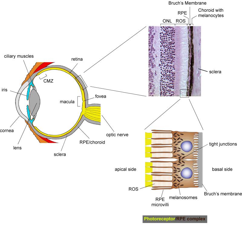
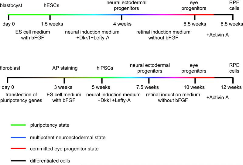
Compared with neural crest-derived melanocytes, retinal pigment epithelium (RPE) cells in the back of the eye are pigment cells of a different kind. They are a part of the brain, form an epithelial monolayer, respond to distinct extracellular signals, and provide functions that far exceed those of a light-absorbing screen. For instance, they control nutrient and metabolite flow to and from the retina, replenish 11-cis-retinal by re-isomerizing all-trans-retinal generated during photoconversion, phagocytose daily a portion of the photoreceptors' outer segments, and secrete cytokines that locally control the innate and adaptive immune systems. Not surprisingly, RPE cell damage is a major cause of human blindness worldwide, with age-related macular degeneration a prevalent example. RPE replacement therapies using RPE cells generated from embryonic or induced pluripotent stem cells provide a novel approach to a rational treatment of such forms of blindness. In fact, RPE-like cells can be obtained relatively easily when stem cells are subjected to a two-step induction protocol, a first step that leads to a neuroectodermal fate and a second to RPE differentiation. Here, we discuss the characteristics of such cells, propose criteria they should fulfill in order to be considered authentic RPE cells, and point out the challenges one faces when using such cells in attempts to restore vision.
John Wiley & Sons A/S. This article is a US Government work and is in the public domain in the USA.
Figures
References
-
- Araki M. Regeneration of the amphibian retina: role of tissue interaction and related signaling molecules on RPE transdifferentiation. Dev. Growth Differ. 2007;49:109–120. - PubMed
-
- Baird PN, Richardson AJ, Robman LD, Dimitrov PN, Tikellis G, McCarty CA, Guymer RH. Apolipoprotein (APOE) gene is associated with progression of age-related macular degeneration (AMD) Hum. Mutat. 2006;27:337–342. - PubMed
Publication types
MeSH terms
Grants and funding
LinkOut - more resources
Full Text Sources
Other Literature Sources
Medical
