

Hif1α down-regulation is associated with transposition of great arteries in mice treated with a retinoic acid antagonist
- PMID: 20846364
- PMCID: PMC2996993
- DOI: 10.1186/1471-2164-11-497
Hif1α down-regulation is associated with transposition of great arteries in mice treated with a retinoic acid antagonist
Abstract
Background: Congenital heart defect (CHD) account for 25% of all human congenital abnormalities. However, very few CHD-causing genes have been identified so far. A promising approach for the identification of essential cardiac regulators whose mutations may be linked to human CHD, is the molecular and genetic analysis of heart development. With the use of a triple retinoic acid competitive antagonist (BMS189453) we previously developed a mouse model of congenital heart defects (81%), thymic abnormalities (98%) and neural tube defects (20%). D-TGA (D-transposition of great arteries) was the most prevalent cardiac defect observed (61%). Recently we were able to partially rescue this abnormal phenotype (CHD were reduced to 64.8%, p = 0.05), by oral administration of folic acid (FA). Now we have performed a microarray analysis in our mouse models to discover genes/transcripts potentially implicated in the pathogenesis of this CHD.
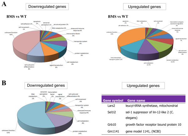
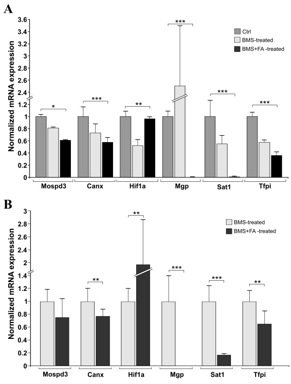
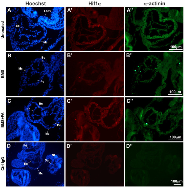
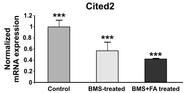
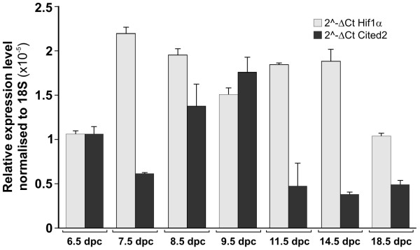
Results: We analysed mouse embryos (8.5 dpc) treated with BMS189453 alone and with BMS189453 plus folic acid (FA) by microarray and qRT-PCR. By selecting a fold change (FC) ≥ ± 1.5, we detected 447 genes that were differentially expressed in BMS-treated embryos vs. untreated control embryos, while 239 genes were differentially expressed in BMS-treated embryos whose mothers had also received FA supplementation vs. BMS-treated embryos. On the basis of microarray and qRT-PCR results, we further analysed the Hif1α gene. In fact Hif1α is down-regulated in BMS-treated embryos vs. untreated controls (FCmicro = -1.79; FCqRT-PCR = -1.76; p = 0.005) and its expression level is increased in BMS+FA-treated embryos compared to BMS-treated embryos (FCmicro = +1.17; FCqRT-PCR = +1.28: p = 0.005). Immunofluorescence experiments confirmed the under-expression of Hif1α protein in BMS-treated embryos compared to untreated and BMS+FA-treated embryos and, moreover, we demonstrated that at 8.5 dpc, Hif1α is mainly expressed in the embryo heart region.
Conclusions: We propose that Hif1α down-regulation in response to blocking retinoic acid binding may contribute to the development of cardiac defects in mouse newborns. In line with our hypothesis, when Hif1α expression level is restored (by supplementation of folic acid), a decrement of CHD is found. To the best of our knowledge, this is the first report that links retinoic acid metabolism to Hif1α regulation and the development of D-TGA.
Figures
References
-
- Jing-Bin H, Ying-Long L, Pei-Wu S, Xiao-Dong L, Ming D, Xiang-Ming F. Molecular mechanisms of congenital heart disease. Cardiovasc Pathol. in press Corrected Proof, Available online 10 September 2009. - PubMed
MeSH terms
Substances
LinkOut - more resources
Full Text Sources
Molecular Biology Databases
