. 2010 Sep 16:11:119.
doi: 10.1186/1471-2202-11-119.

The effect of mesenchymal stem cell transplantation on the recovery of bladder and hindlimb function after spinal cord contusion in rats

Affiliations

The effect of mesenchymal stem cell transplantation on the recovery of bladder and hindlimb function after spinal cord contusion in rats

Won Beom Park et al. BMC Neurosci. .

Abstract

Background: Mesenchymal stem cells are widely used for transplantation into the injured spinal cord in vivo model and for safety, many human clinical trials are continuing to promote improvements of motor and sensory functions after spinal cord injury. Yet the exact mechanism for these improvements remains undefined. Neurogenic bladder following spinal cord injury is the main problem decreasing the quality of life for patients with spinal cord injury, but there are no clear data using stem cell transplantation for the improvement of neurogenic bladder for in vivo studies and the clinical setting.The purpose of this study was to delineate the effect of human mesenchymal stem cell (hMSCs) transplantation on the restoration of neurogenic bladder and impaired hindlimb function after spinal cord contusion of rats and the relationship between neurotrophic factors such as brain derived neurotrophic factor (BDNF) and neurotrophin-3 (NT-3) and bladder and hindlimb functions.

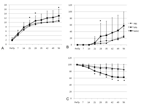
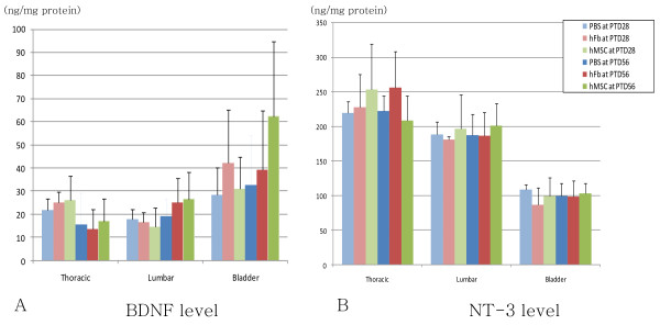
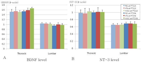
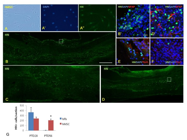
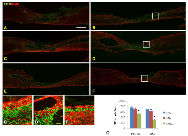
Results: Modified moderate contusion injury were performed on the thoracic spinal cord of Sprague-Dawley rats using MASCIS impactor and hMSCs, human fibroblasts or phosphate-buffered saline were transplanted into injured spinal cord 9 days after injury for hMSC and two control groups respectively. Ladder test showed more rapid restoration of hindlimb function in hMSC group than in control group, but Basso, Beattie, and Bresnahan score and coupling score were not different significantly among hMSC and two control groups. Neurogenic bladder was not improved in either group. ED1 positive macrophages were significantly reduced in hMSC group than in two control groups, but ELISA and RT-PCR studies revealed BDNF and NT-3 levels in spinal cord and bladder were not different among hMSC and two control groups regardless the experimental duration.

Conclusion: hMSC transplantation was effective in reducing inflammatory reaction after spinal cord contusion of rats but not sufficient to recover locomotor and bladder dysfunction. BDNF and NT-3 levels in the spinal cord and bladder were not increased 28 and 56 days after hMSC transplantation.


Figures

Figure 1
Figure 1
Locomotor function in experimental (hMSC) and two control (PBS and hFb) groups. (A) BBB (Basso, Beattie, Bresnahan) scores, (B) coupling scores and (C) ladder scores showed gradual restoration in experimental and control groups, but all three scales of experimental group (hMSC) at some periods (asterisks) were restored more than those of control group (hFb). PTD: post-transplantation day, PBS: control group received phosphate-buffered saline, hFb: control group received human fibroblast, hMSC: experimental group received human mesenchymal stem cell, *p < 0.05 by one-way ANOVA with Bonferroni post hoc test.
Figure 2
Figure 2
mRNA levels of neurotrophic factors by ELISA in experimental (hMSC) and two control (PBS and hFb) groups at 28 and 56 days after transplantation. (A) BDNF level and (B) NT-3 level in the thoracic and lumbar spinal cords and bladder showed no statistical difference among experimental and two control groups at PTD28 and PTD56. hMSC: experimental group received human mesenchymal stem cell, PBS: control group received phosphate-buffered saline, hFb: control group received human fibroblast, PTD: post-transplantation day
Figure 3
Figure 3
Protein levels of neurotrophic factors by RT-PCR in experimental (hMSC) and two control (PBS and hFb) groups at 28 and 56 days after transplantation. (A) BDNF level and (B) NT-3 level in the thoracic and lumbar spinal cords also showed no statistical difference among experimental and two control groups at PTD28 and PTD56. hMSC: experimental group received human mesenchymal stem cell, PBS: control group received phosphate-buffered saline, hFb: control group received human fibroblast, PTD: post-transplantation day
Figure 4
Figure 4
Immunohistochemistry of human mesenchymal stem cells (hMSCs) in vitro and in vivo studies. (A) hMSCs were well stained by anti-human nucleus antibody (HN) in vitro (A"), and injured area of thoracic spinal cord after transplantation of human fibroblasts (hFbs) at PTD28 (B) and after transplantation of hMSCs at PTD28 (C) and PTD56 (D) was stained with HN. B' and D' images are magnified images of B and D white-lined boxes respectively. Double staining with HN (green color) and anti-GFAP antibody (red color) (B' and D'), and HN (green color) and anti-beta III tubulin antibody (red color) after hMSC transplantation at PTD28 (E) and PTD56 (F) indicated that transplanted hMSCs into injured spinal cord did not differentiated into neurons nor astrocytes 28 and 56 days after transplantation. (G) Total number of HN-positive cells per section was higher in hMSC group than in hFb group (asterisk) at PTD56. White scale bar in B = 500 μm, yellow scale bar in B' = 20 μm.
Figure 5
Figure 5
Injured spinal cords of PBS, hFB and hMSC groups at PTD28 and PTD56 stained with anti-macrophages/monocytes (ED1) antibody (green color) and anti-GFAP antibody (red color; A-F). PBS group at PTD28 (A) and PTD56 (B), and hFB group at PTD28 (C) and PTD56 (D) showed many macropahges and monocytes were existed within the injured spinal cords compared with hMSC group at PTD28 (E) and PTD56 (F). . B', D' and F' images are magnified images of B, D and F white-lined boxes respectively. The number of macrophages and monocytes was lower in hMSC group than in PBS and hFb groups at PTD28 and PTD56 (asterisk, G). White scale bar in A = 1 mm, yellow scale bar in D' = 100 μm.

References

    1. Ku JH. The management of neurogenic bladder and quality of life in spinal cord injury. BJU Int. 2006;11:739–745. doi: 10.1111/j.1464-410X.2006.06395.x. - DOI - PubMed
    1. Garcia Leoni ME, Esclarin De Ruz A. Management of urinary tract infection in patients with spinal cord injuries. Clin Microbiol Infect. 2003;11:780–785. doi: 10.1046/j.1469-0691.2003.00643.x. - DOI - PubMed
    1. Shoichet MS, Tator CH, Poon P, Kang C, Baumann MD. Intrathecal drug delivery strategy is safe and efficacious for localized delivery to the spinal cord. Prog Brain Res. 2007;11:385–392. full_text. - PubMed
    1. Nomura H, Tator CH, Shoichet MS. Bioengineered strategies for spinal cord repair. J Neurotrauma. 2006;11:496–507. doi: 10.1089/neu.2006.23.496. - DOI - PubMed
    1. Willerth SM, Sakiyama-Elbert SE. Cell therapy for spinal cord regeneration. Adv Drug Deliv Rev. 2008;11:263–276. doi: 10.1016/j.addr.2007.08.028. - DOI - PMC - PubMed

Publication types

MeSH terms