. 2010 Sep 17:9:249.
doi: 10.1186/1476-4598-9-249.

Molecular detection, quantification, and isolation of Streptococcus gallolyticus bacteria colonizing colorectal tumors: inflammation-driven potential of carcinogenesis via IL-1, COX-2, and IL-8

Affiliations

Molecular detection, quantification, and isolation of Streptococcus gallolyticus bacteria colonizing colorectal tumors: inflammation-driven potential of carcinogenesis via IL-1, COX-2, and IL-8

Ahmed S Abdulamir et al. Mol Cancer. .

Abstract

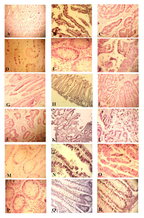
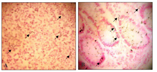
Background: Colorectal cancer (CRC) has long been associated with bacteremia and/or endocarditis by Streptococcus gallolyticus member bacteria (SGMB) but the direct colonization of SGMB along with its molecular carcinogenic role, if any, has not been investigated. We assessed the colonization of SGMB in CRC patients with history of bacteremia (CRC-w/bac) and without history of bacteremia (CRC-wo/bac) by isolating SGMB from feces, mucosal surfaces of colorectum, and colorectal tissues and detecting SGMB DNA, via PCR and in situ hybridization (ISH) assays targeting SodA gene in colorectal tissues. Moreover, mRNA of IL1, IL-8, COX-2, IFN-γ, c-Myc, and Bcl-2 in colorectal tissues of studied groups was assessed via ISH and RT-PCR.

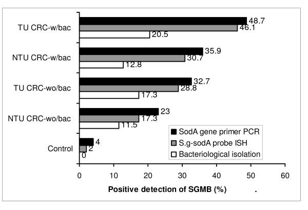
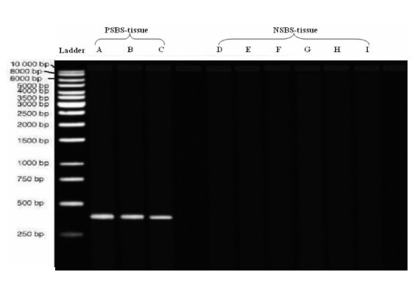
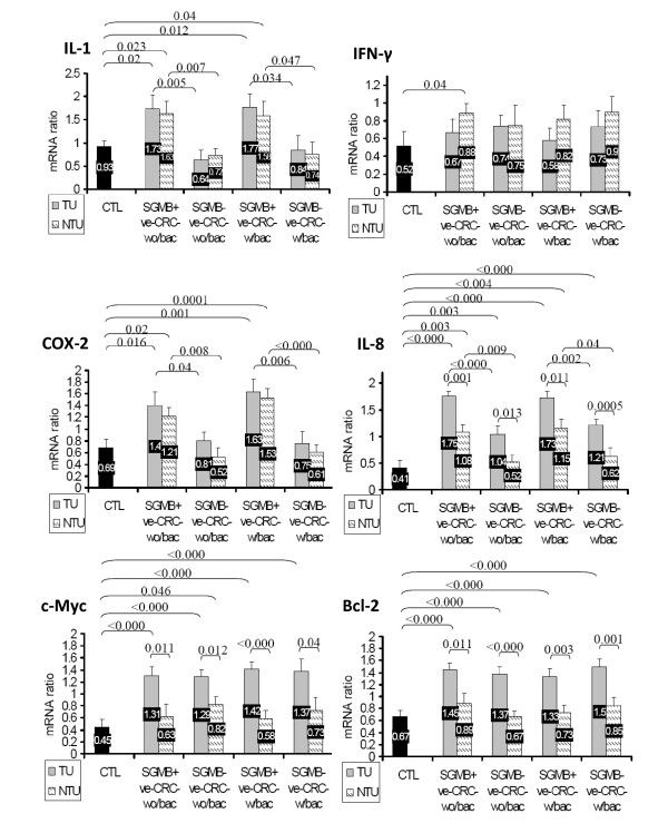
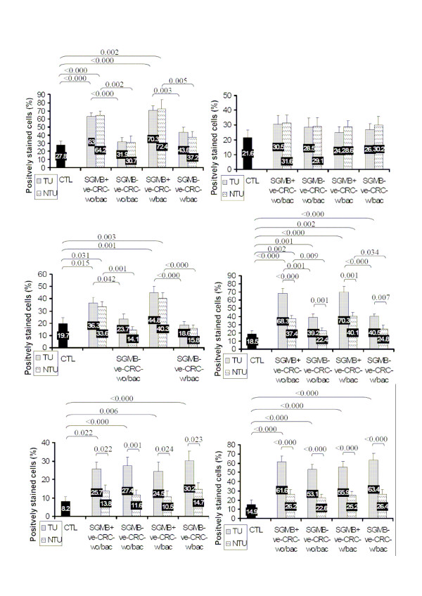
Results: SGMB were found to be remarkably isolated in tumorous (TU) and non-tumorous (NTU) tissues of CRC-w/bac, 20.5% and 17.3%, and CRC-wo/bac, 12.8% and 11.5%, respectively while only 2% of control tissues revealed SGMB (P < 0.05); such contrast was not found in mucosal and fecal isolation of SGMB. The positive detection of SGMB DNA in TU and NTU of CRC-w/bac and CRC-wo/bac via PCR, 48.7%, 35.9%, 32.7%, and 23%, respectively, and ISH, 46.1%, 30.7%, 28.8%, and 17.3%, respectively, was higher than in control tissues, 4 and 2%, respectively (P < 0.05). SGMB count measured via quantitative PCR of SGMB DNA in terms of copy number (CN), in TU and NTU of CRC-w/bac and CRC-wo/bac, 2.96-4.72, 1.29-2.81, 2.16-2.92, and 0.67-2.07 log10 CN/g respectively, showed higher colonization in TU than in NTU and in CRC-w/bac than in CRC-wo/bac (P < 0.05). The PCR-based mRNA ratio and ISH-based percentage of positively stained cells of IL-1, 1.77 and 70.3%, COX-2, 1.63 and 44.8%, and IL-8, 1.73 and 70.3%, respectively, rather than IFN-γ, c-Myc, and Bcl-2, were higher in SGMB positive patients than in control or SGMB negative patients (P < 0.05).

Conclusions: The current study indicated that colorectal cancer is remarkably associated with SGMB; moreover, molecular detection of SGMB in CRC was superior to link SGMB with CRC tumors highlighting a possible direct and active role of SGMB in CRC development through most probably inflammation-based sequel of tumor development or propagation via, but not limited to, IL-1, COX-2, and IL-8.


Figures

Figure 1
Figure 1
A histogram showing a comparison in the percentage of the positive detection/isolation of SGMB among control, TU and NTU tissues of CRC-wo/bac, and TU and NTU tissues of CRC-w/bac groups using three detection methods, namely SodA gene primer PCR, S.g-sodA-probe ISH assay, and enrichment-based bacteriological methods. It is shown that the top SGMB detection method was PCR, then ISH assay, and least effective was bacteriological isolation.
Figure 2
Figure 2
Gel electrogram of SodA gene-specific primer-based PCR products of normal colorectal tissues experimentally inoculated with three PSBS strains (PSBS-tissue) and six NSBS strains (NSBS-tissue). PSBS-tissues (lanes A-C) were CIP 105428, CIP 105683, and CIP 105070, respectively while NSBS-tissues (lanes D-I) were CIP 108103, CIP 56.41, CIP 102504, ACM 3539, CIP 76117, and ATCC 25285, respectively. PSBS-tissues, but not NSBS-tissues samples, yielded single band at 408 bp. The used ladder was QIAGEN GelPilot DNA Molecular Weight Marker.
Figure 3
Figure 3
Normalized mRNA ratio, expressed as mean+ SE, of IL-1, IFN-γ, COX-2, IL-8, c-Myc, and Bcl-2 is shown in TU and NTU tissues of control, SGMB+ve-CRC-wo/bac, SGMB-ve-CRC-wo/bac, SGMB+ve-CRC-w/bac and SGMB-ve-CRC-w/bac. Significant differences in mean mRNA ratio (P < 0.05) among different groups are shown as demarcation brace lines with corresponding P values whereas insignificant differences are devoid of demarcation and P values.
Figure 4
Figure 4
Mean+SE of positive mRNA staining via ISH assay for IL-1, IFN-γ, COX-2, IL-8, c-Myc, and Bcl-2 is shown in TU and NTU tissues of control, SGMB+ve-CRC-wo/bac, SGMB-ve-CRC-wo/bac, SGMB+ve-CRC-w/bac and SGMB-ve-CRC-w/bac. Significant differences in mean of positive mRNA staining (P < 0.05) among different groups was shown as demarcation brace lines with corresponding P values whereas insignificant differences were devoid of demarcation and P values.
Figure 5
Figure 5
Examples of ISH staining of mRNA of IL-1, IFN-γ, COX-2, IL-8, c-Myc, and Bcl-2. Cells stained positive are shown with dark color of NBT/BCIP in the nuclei of glandular cells as well as stromal cells. mRNA expression of IL-1 (A-C) at X100, IFN-γ (D-F) at 400X, COX-2 (G-I) at X100, and IL-8 (J-L) at 100X, is shown in control, TU SGMB+ve-CRCw/bac, and TU SGMB-ve-CRC-w/bac, respectively. mRNA expression of c-Myc (M-O) at X400 and Bcl-2 (P-R) at X400 is shown in control, TU SGMB-ve-CRC-wo/bac, and NTU SGMB-ve-CRC-wo/bac.
Figure 6
Figure 6
Examples of ISH staining of SGMB DNA at 400X in colorectal tissues of (A) TU SGMB+ve-CRC-w/bac and (B) NTU SGMB+ve-CRC-wo/bac. SGMB DNA staining was shown as chains of stained tiny dark spots which are pointed by black arrows. No distinctive pattern of distribution of the positively stained SGMB was seen throughout stained sections.

References

    1. Triantafillidis JK, Nasioulas G, Kosmidis PA. Colorectal cancer and inflammatory bowel disease: epidemiology, risk factors, mechanisms of carcinogenesis and prevention strategies. Anticancer Res. 2009;29(7):2727–2737. - PubMed
    1. Ablashi DV, Chatlynne LG, Whitman JE Jr, Cesarman E. Spectrum of Kaposi's sarcoma-associated herpesvirus, or human herpesvirus 8, diseases. Clin Microbiol Rev. 2002;15(3):439–464. doi: 10.1128/CMR.15.3.439-464.2002. - DOI - PMC - PubMed
    1. Boshoff C, Weiss R. AIDS-related malignancies. Nat Rev Cancer. 2002;2(5):373–382. doi: 10.1038/nrc797. - DOI - PubMed
    1. Parsonnet J. Bacterial infection as a cause of cancer. Environ Health Perspect. 1995;103(Suppl 8):263–268. doi: 10.2307/3432323. - DOI - PMC - PubMed
    1. Malfertheiner P, Sipponen P, Naumann M, Moayyedi P, Megraud F, Xiao SD, Sugano K, Nyren O. Helicobacter pylori eradication has the potential to prevent gastric cancer: a state-of-the-art critique. Am J Gastroenterol. 2005;100(9):2100–2115. doi: 10.1111/j.1572-0241.2005.41688.x. - DOI - PubMed