. 2010 Sep 17;17(1):76.
doi: 10.1186/1423-0127-17-76.

Suberoylanilide hydroxamic acid induces apoptosis and sub-G1 arrest of 320 HSR colon cancer cells

Affiliations

Suberoylanilide hydroxamic acid induces apoptosis and sub-G1 arrest of 320 HSR colon cancer cells

Pei-Chang Sun et al. J Biomed Sci. .

Abstract

Background: Histone deacetylases and histone acetyl transferases covalently modify histone proteins, consequentially altering chromatin architecture and gene expression.

Methods: The effects of suberoylanilide hydroxamic acid, a HDAC inhibitor, on 320 HSR colon cells were assessed in 320 HSR colon cancer cells.

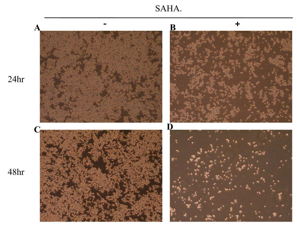
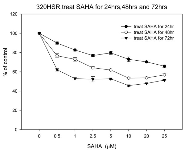
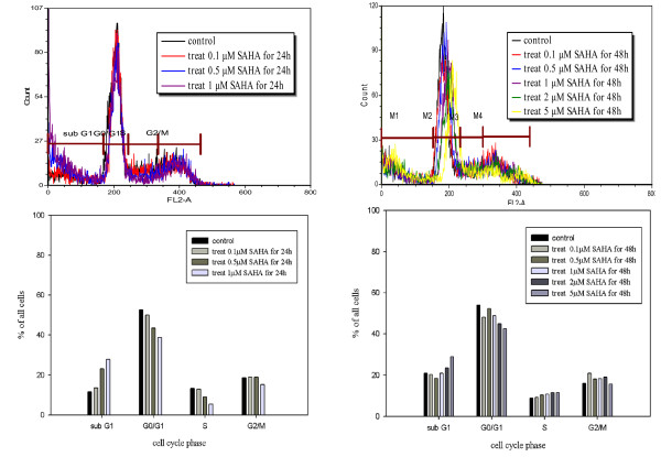
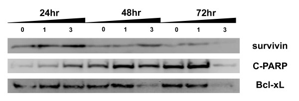
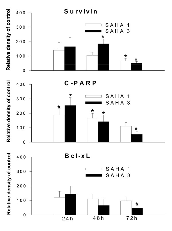
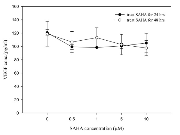
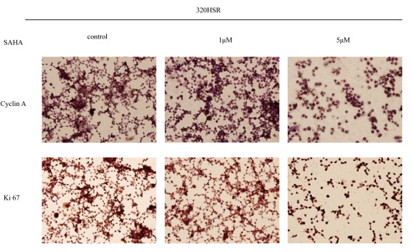
Results: Concentration and time-dependent inhibition of 320 HSR cell proliferation was observed. Treatment of 320 HSR cells with 5 μM SAHA for 72 h significantly inhibited their growth by 50% as compared to that of the control. Fluorescence-activated cell sorting analysis demonstrated significant inhibition of cell cycle progression (sub-G1 arrest) and induction of apoptosis upon various SAHA concentrations after 48 h. In addition, the anti-apoptosis proteins, survivin and Bcl-xL, were significantly inhibited by SAHA after 72 h of treatment. Immunocytochemistry analysis revealed that SAHA-resistant cells were positive for cyclin A (85%), ki-67 (100%), p53 (100%), survivin (100%), and p21 (90%) expression. Furthermore, a significant increase cyclin A-, Ki-67-, p53-, survivin-, and p21-positive cells were noted in SAHA-resistant tumor cells.

Conclusion: Our results demonstrated for the first time in 320 HSR colon adenocarcinoma cells that SAHA might be considered as an adjuvant therapy for colon adenocarcinoma.


Figures

Figure 1
Figure 1
Morphology of 320 HSR colon cancer cells after 48 h treatment with 5 μM SAHA. Original magnification ×400.
Figure 2
Figure 2
Effects of SAHA on 320 HSR cell growth using the MTT assay. 320 HSR cells were treated with various concentrations of SAHA for 24, 48, and 72 h.
Figure 3
Figure 3
SAHA induces apoptosis in colon cancer cell lines. 320 HSR cells were stained with Annexin V (FITC) and propidium iodide (PI) after treatment with SAHA. Fluorescence-activated cell sorting analysis of 320 HSR cancer cell line at 48 h following treatment with 0, 1, 2.5, and 5 μM SAHA (A, B, C, D, respectively). Percentages represent Annexin V-positive/PI-negative (early apoptotic) and Annexin V-positive/PI-positive cells (apoptotic).
Figure 4
Figure 4
Fluorescence activated cell sorting (FACS) analysis revealed SAHA-induced sub-G1 arrest in 320HSR cells. Cells were harvested 48 h after stimulation in the absence or presence of SAHA (0.1 μM to 5 μM). Intracellular PI fluorescence intensities of cells are presented in the upper panels. The percentage of cells in the G0/G1 phase was significantly inhibited by SAHA treatment after 24 or 48 h. The percentage of cells in the sub-G1 phase was significantly increased in response to SAHA treatment.
Figure 5
Figure 5
SAHA alters survivin, C-PARP, and Bcl-xL protein levels in 320 HSR cells. Western blot analysis of cells treated with or without SAHA (1 or 3 μM) for 24, 48, and 72 h.
Figure 6
Figure 6
Histogram values are means and standard error of band densitometry data measured in figure 5. *P <0.05 vs. control.
Figure 7
Figure 7
Effects of SAHA on VEGF secretion by 320 HSR cells. Cells were treated with different SAHA concentrations for 24 h and 48 h. VEGF concentrations in the conditioned medium were determined by ELISA and normalized to 1 × 10⁵ viable cells.
Figure 8
Figure 8
Immunocytochemical analysis of cyclin A and Ki-67 expression in 320 HSR cells. Cells were treated with or without SAHA (1 or 5 μM) for 48 h. Original magnification ×400.
Figure 9
Figure 9
Immunocytochemical analysis of p53, survivin, and p21 expression in 320 HSR cells. Cells were treated with or without SAHA (1 or 5 μM) for 48 h. Original magnification ×400.

References

    1. Ito K, Adcock IM. Histone acetylation and histone deacetylation. Mol Biotechnol. 2002;20:99–106. doi: 10.1385/MB:20:1:099. - DOI - PubMed
    1. Kuo MH, Allis CD. Roles of histone acetyltransferases and deacetylases in gene regulation. Bioassays. 1998;20:615–626. doi: 10.1002/(SICI)1521-1878(199808)20:8<615::AID-BIES4>3.0.CO;2-H. - DOI - PubMed
    1. Marks PA, Richon VM, Breslow R, Rifkind RA. Histone deacetylase inhibitors as new cancer drugs. Curr Opin Oncol. 2001;13:477–483. doi: 10.1097/00001622-200111000-00010. - DOI - PubMed
    1. Kim DH, Kim M, Kwon HJ. Histone deacetylase in carcinogenesis and its inhibitors as anti-cancer agents. J Biochem Mol Biol. 2003;36:110–119. - PubMed
    1. Truchet I, Jozan S, Baron S, Frongia C, Balaguer P, Richard-Foy H, Valette A. Estrogen and antiestrogen-dependent regulation of breast cancer cell proliferation in multicellular spheroids: Influence of cell microenvironment. Int J Oncol. 2008;32:1033–1039. - PubMed

Publication types

MeSH terms