. 2011 Jan;39(2):623-34.
doi: 10.1093/nar/gkq802. Epub 2010 Sep 15.

Mutational and biochemical analysis of the DNA-entry nuclease EndA from Streptococcus pneumoniae

Affiliations

Mutational and biochemical analysis of the DNA-entry nuclease EndA from Streptococcus pneumoniae

Marika Midon et al. Nucleic Acids Res. 2011 Jan.

Abstract

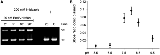
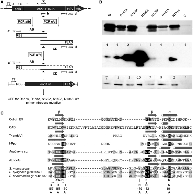
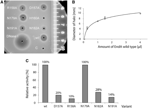
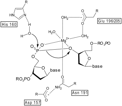
EndA is a membrane-attached surface-exposed DNA-entry nuclease previously known to be required for genetic transformation of Streptococcus pneumoniae. More recent studies have shown that the enzyme also plays an important role during the establishment of invasive infections by degrading extracellular chromatin in the form of neutrophil extracellular traps (NETs), enabling streptococci to overcome the innate immune system in mammals. As a virulence factor, EndA has become an interesting target for future drug design. Here we present the first mutational and biochemical analysis of recombinant forms of EndA produced either in a cell-free expression system or in Escherichia coli. We identify His160 and Asn191 to be essential for catalysis and Asn182 to be required for stability of EndA. The role of His160 as the putative general base in the catalytic mechanism is supported by chemical rescue of the H160A variant of EndA with imidazole added in excess. Our study paves the way for the identification and development of protein or low-molecular-weight inhibitors for EndA in future high-throughput screening assays.


Figures

Figure 1.
Figure 1.
(A) SDS–PAGE analysis of the expression of EndA H160A fused to the pelB leader sequence for periplasmic localization in E. coli. (u) cells before induction, suspended in loading buffer (i) cells after induction, suspended in loading buffer; (med-u) concentrated culture medium before induction, (med-i) concentrated culture medium after induction, (wcl-i) whole cell lysate after cell disruption by sonication and (sf-i) soluble fraction after centrifugation of the whole cell lysate. (B) Purification of His-GST double-tagged EndA H160A. Upper panel: SDS–PAGE analysis of the lysate of transformed BL21-Gold(DE3) cells before (u) and after (i) induction; (IMAC-IDA) His-GST-EndA H160A purified via immobilized metal ion affinity chromatography using a Ni2+-IDA-column; (TEV) cleavage of His-GST-EndA H160A with TEV protease; (IMAC-NTA ft) flow through after incubation with Ni2+-NTA-Agarose; (SEC) EndA-H160A after size-exclusion chromatography. Lower panel: western blot using anti-GST antibody to monitor the purification steps described above. (C) Purification of EndA H160A by size-exclusion chromatography: after clipping off the His-GST double tag and incubation with Ni-NTA-Agarose the collected flow through containing only untagged EndA H160A was applied to a Superdex 75 column and eluted at a flow rate of 1 ml/min. The black triangle indicates the time point of sample loading. Elution of proteins was monitored at 280 nm. Peak I represents EndA separated from a residual contaminant (II). (D) Retention coefficients (Superdex 75 column) of EndA and protein standards were determined and plotted against the natural logarithm of the molecular mass. The molecular weight of 25.5±4 kDa obtained for EndA, based on the fitted curve, corresponds reasonably well with the calculated molecular weight of 26.8 kDa indicating that EndA is a monomer under these conditions.
Figure 2.
Figure 2.
SAXS of EndA. (A) SAXS intensity I(q) data for EndA H160A (3.0 mg/ml); the insets show the P(r) pair-wise vector length distribution curve and Guinier fit of the I(q) scattering data. The gray line in the I(q) scattering plot is the calculated scattering curve of NucA generated with CRYSOL. (B) Ab-initio models derived from SAXS intensity data (surface representation) superimposed with a ribbon representation of the Anabaena NucA crystal structure (pdb-code 1zm8).
Figure 3.
Figure 3.
Chemical rescue of the nuclease activity of the catalytically inactive EndA His160A variant by imidazole. (A) Aliquots of the reaction mixture containing 20 nM EndA H160A and 15 ng/µl pBSK plasmid in triple buffer at pH 8.0 were taken at the time points indicated and the reaction products analyzed by 0.8% agarose gel electrophoresis. The analysis shows an increase of open circular (oc) and a decrease of supercoiled (sc) plasmid DNA over time. Plasmid DNA incubated for 20 min in the absence of EndA H160A and plasmid (C) were loaded as controls. (B) The experiment was repeated at different pH-values (pH 5.5–9.5) and the ratio of open circular to super-coiled plasmid DNA plotted against time (n = 3). EndA H160A showed nucleolytic activity at slightly basic pH values ranging from pH 7.5 to 8.5.
Figure 4.
Figure 4.
(A) Generation of template DNA for in vitro protein synthesis via overlap extension PCR. Plasmid pET25+ harboring the gene coding for the inactive EndA H160A variant was used as a template for overlap extension PCR to generate the gene for wild-type EndA. The endA H160A gene was segmented via amplification in two separate PCR reactions with primer a containing a ribosomal binding site and primer b for segment AB, and primers c and d containing the sequence coding for the FLAG tag in segment CD. The full-length gene was generated by using primer a′ containing a T7 promoter sequence and primer d. Replacement of Ala160 by histidine was achieved by the two internal primer b and c introducing the nucleotide substitution (black triangle) within the overlap of the gene segments. The PCR product was the template for in vitro synthesis of wild-type EndA and for overlap extension PCR to produce the EndA variants D157A, R158A, N179A, N182A and N191A using primer c and d to introduce the mutations. The gene for the variant H160A was generated with primer a and d and in a second PCR extended with the T7 promoter sequence with primer a′, using d as reverse primer. (B) In-gel activity assay and western blot analysis of wild-type EndA and its variants. Upper panel: nuclease activity of the crude in vitro protein synthesis reaction mixture of wild-type EndA and variants D157A, R158A, H160A, N179A, N182A and N191A separated on an SDS–PAGE gel containing high-molecular-weight herring sperm DNA (ethidium bromide stained). The reaction mixture with the control template supplied with the In vitro Protein Synthesis Kit (New England BioLabs) was used as a negative control (C). Dark bands correspond to gel areas with digested DNA that are not stained with ethidium bromide. Middle and lower panel: western blot analysis of FLAG-tagged EndA and variants D157A, R158A, H160A, N179A, N182A, N191A using an anti-FLAG antibody. Numbers indicate the amounts (µl) of the respective crude synthesis reaction mixture loaded onto the gel. (C) Alignment of DNA-entry nucleases with other ββα-Me-finger enzymes (H-N-H/N- and DRGH-motif containing nucleases) of known structure. Indicated are amino acid residues selected for a substitution by alanine in S. pneumoniae EndA. Mutations at positions 157, 158 and 160 concern the conserved DRGH-motif. Histidine at position 160 also represents the first amino acid within the H-N-H/N-motif and plays a central role in catalysis. One of two asparagines at positions 179 and 182 could represent the first asparagine residue of the H-N-H/N-motif, with asparagine 191 being the second. The alignment was manually refined based on the structures of the listed enzymes and our mutational analysis (Colicin E9, pdb code 1emv; Caspase-activated DNase (CAD), pdb-code 1v0d; T4 endonuclease VII, pdb code 1e7d; I-PpoI, pdb-code 1a74; Anabaena nuclease (NucA), pdb code 1zm8; Endonuclease G (dEndoG) from D. melanogaster, pdb code 3ism; Serratia nuclease, pdb code 1smn). Secondary structure elements forming the core of the ββα-Me-finger active-site motif are indicated by arrows (β-sheets) and tubes (α-helices) as derived from the respective crystal structures. For the DNA-entry nucleases from S. pyogenes and S. pneumoniae (EndA) the gi numbers are indicated.
Figure 5.
Figure 5.
SRED assay: (A) Relative nuclease activity of the in vitro protein synthesis reaction mixture containing wild-type EndA, D157A, R158A, N179A, N182A and N191A tested with high-molecular-weight herring sperm DNA embedded in 1% agarose (prestained with ethidium bromide), 40 mM Tris–HCl, pH 7.7, 2 mM MgCl2. Variant H160A showed no measurable activity in this assay. The reaction mixture with the control template supplied with the in vitro Protein Synthesis Kit (New England BioLabs) was used as a negative control (C), DNaseI (Fermentas) as positive control. The ruler indicates the diameter of the halos. (B) Calibration curve for the estimation of relative nucleolytic activity from the size of the halos. The size of the halos (diameter in millimeters) was plotted against the amount (µl) of the protein synthesis reaction mixture containing wild-type EndA. (C) The relative nucleolytic activity of each EndA variant contained in the crude reaction mixture was determined from the diameter of the halos using the calibration curve in (B) and the concentration of the EndA variants in the crude reaction mixture determined by western blot analysis. The activity of wild-type EndA is set to 100%.
Figure 6.
Figure 6.
Substrate preference of wild-type EndA: Nucleolytic activity of wild-type EndA was tested on (A) ΦX174 DNA (single-stranded, circular), (B) RF I DNA (double-stranded, circular) and (C) RF II DNA (double-stranded, nicked). Wild-type EndA shows no substrate preference (ss—single stranded, sc—super coiled, oc—open circle, li—linear).
Figure 7.
Figure 7.
Catalytic mechanism of wild-type EndA: the proposed catalytic mechanism of EndA is based on the established catalytic mechanism of the Serratia nuclease (26). His160 acts as the general base for the activation of a water molecule. The magnesium ion is coordinated by Asn191, which is held in place by Asp157, and the proS-oxygen as well as the 3′-oxygen of the phosphate diester. A glutamic acid residue residue nearby (Glu196 or Glu205 in EndA) is assumed to be indirectly involved in water-mediated magnesium ion binding.

References

    1. Lacks S, Neuberger M. Membrane location of a deoxyribonuclease implicated in the genetic transformation of Diplococcus pneumoniae. J. Bacteriol. 1975;124:1321–1329. - PMC - PubMed
    1. Puyet A, Greenberg B, Lacks SA. Genetic and structural characterization of endA. A membrane-bound nuclease required for transformation of Streptococcus pneumoniae. J. Mol. Biol. 1990;213:727–738. - PubMed
    1. Buchanan JT, Simpson AJ, Aziz RK, Liu GY, Kristian SA, Kotb M, Feramisco J, Nizet V. DNase expression allows the pathogen group A Streptococcus to escape killing in neutrophil extracellular traps. Curr. Biol. 2006;16:396–400. - PubMed
    1. Beiter K, Wartha F, Albiger B, Normark S, Zychlinsky A, Henriques-Normark B. An endonuclease allows Streptococcus pneumoniae to escape from neutrophil extracellular traps. Curr. Biol. 2006;16:401–407. - PubMed
    1. Brinkmann V, Reichard U, Goosmann C, Fauler B, Uhlemann Y, Weiss DS, Weinrauch Y, Zychlinsky A. Neutrophil extracellular traps kill bacteria. Science. 2004;303:1532–1535. - PubMed

Publication types

MeSH terms