. 2010 Sep 17;329(5998):1492-9.
doi: 10.1126/science.1188015.

Evidence for an alternative glycolytic pathway in rapidly proliferating cells

Affiliations

Evidence for an alternative glycolytic pathway in rapidly proliferating cells

Matthew G Vander Heiden et al. Science. .

Abstract

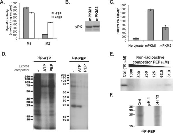
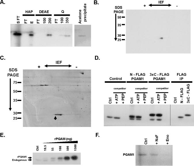
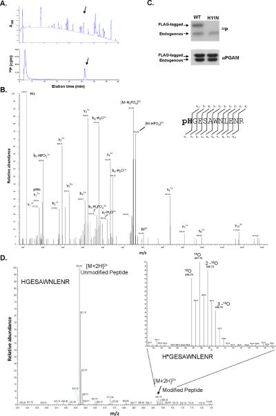
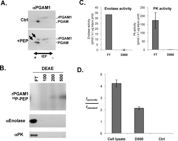
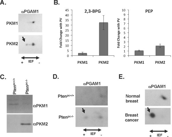
Proliferating cells, including cancer cells, require altered metabolism to efficiently incorporate nutrients such as glucose into biomass. The M2 isoform of pyruvate kinase (PKM2) promotes the metabolism of glucose by aerobic glycolysis and contributes to anabolic metabolism. Paradoxically, decreased pyruvate kinase enzyme activity accompanies the expression of PKM2 in rapidly dividing cancer cells and tissues. We demonstrate that phosphoenolpyruvate (PEP), the substrate for pyruvate kinase in cells, can act as a phosphate donor in mammalian cells because PEP participates in the phosphorylation of the glycolytic enzyme phosphoglycerate mutase (PGAM1) in PKM2-expressing cells. We used mass spectrometry to show that the phosphate from PEP is transferred to the catalytic histidine (His11) on human PGAM1. This reaction occurred at physiological concentrations of PEP and produced pyruvate in the absence of PKM2 activity. The presence of histidine-phosphorylated PGAM1 correlated with the expression of PKM2 in cancer cell lines and tumor tissues. Thus, decreased pyruvate kinase activity in PKM2-expressing cells allows PEP-dependent histidine phosphorylation of PGAM1 and may provide an alternate glycolytic pathway that decouples adenosine triphosphate production from PEP-mediated phosphotransfer, allowing for the high rate of glycolysis to support the anabolic metabolism observed in many proliferating cells.


Figures

Fig. 1
Fig. 1. Evidence of PEP-dependent phosphorylation of a 25-kD protein in PKM2 expressing cells with less pyruvate kinase activity
A. 6xHis-tagged human PKM1 and PKM2 were expressed in E. coli and purified by Ni-affinity chromatography. The specific activity of each enzyme was determined in the presence of saturating amounts of PEP and ADP. The activity of PKM1 and PKM2 in the presence and absence of FBP is shown. B. H1299 cells were engineered to express equivalent amount of PKM1 or PKM2 protein as described previously (4). Equivalent expression of PKM1 and PKM2 was confirmed by Western blot using an antibody (αPK) that recognizes an epitope shared by PKM1 and PKM2 as shown. C. As in (A), pyruvate kinase activity was determined using saturating amounts of PEP and ADP. The relative pyruvate kinase activity observed in the PKM1- or PKM2-expressing cells described in (B), relative to lysis buffer alone, is shown. D. HEK293 cells were hypotonically lysed and incubated with 32P-labeled ATP or 32P-labeled PEP prior to analysis by SDS-PAGE and autoradiography. The lysates were incubated with 32P-labeled ATP or 32P-labeled PEP in the presence of 10 μM ATP or PEP respectively (−), or with the addition of 1 mM non-radioactive competitor ATP or PEP as shown. E. Cell lysate was incubated with 32P-labeled PEP in the presence of the indicated concentration of non-radioactive competitor PEP prior to analysis by SDS-PAGE and autoradiography as shown. F. Cell lysate was incubated with 32P-labeled PEP as above and the pH of the reaction was changed to pH 1 or pH 13 as indicated. Reactions were incubated for 2 hours at 65° C prior to analysis by SDS-PAGE and autoradiography as shown.
Fig. 2
Fig. 2. PGAM1 as the target of PEP-dependent phosphorylation through an enolase-independent reaction
A. The S100 fraction from a HEK293 cell lysates was passed sequentially through a custom column and a strong cation exchange column prior to incubating with 32P-labeled PEP (S FT). This reaction was then applied to a hydroxyapatite (HAP) column and eluted with 50 mM NaHPO4. The salt elution (E) containing the 32P-labeled species was diluted to < 25 mM NaHPO4 and applied to a weak anion exchange (DEAE) column. Elution from the DEAE column was performed with 100 mM and 200 mM NaCl as indicated. The 200 mM salt fraction containing the 32P-labeled species was diluted to 50 mM NaCl and applied to a strong anion exchange (Q) column and eluted sequentially with 100 mM and 350 mM NaCl as indicated. The 350 mM salt fraction containing the 32P-labeled species was acetone precipitated for analysis by 2D-IEF/SDS-PAGE. An aliquot of each fraction was analyzed by SDS-PAGE and autoradiography as shown. Flow through fractions are indicated as (FT). B. The acetone-precipitated 350 mM salt fraction described in (A) was separated by 2D-IEF and SDS-PAGE and the 32P-labeled species was identified by autoradiography as shown. C. The acetone-precipitated 350 mM salt fraction prepared as described in (A) was separated by 2D-IEF and SDS-PAGE and proteins identified by coomassie stain as shown. The species corresponding to the 32P-labeled species is indicated with an arrow. D. HEK293 cells were transiently transfected with control plasmid (Control), a N-terminally FLAG tagged PGAM1 cDNA (N-FLAG PGAM1), or a C-terminal triple FLAG tagged PGAM1 cDNA (3×C – FLAG PGAM1) as indicated. Hypotonic lysates from these cells were incubated with 32P-labeled PEP alone (Ctrl) or in the presence of 1 mM cold competitor ATP or PEP as indicated. The products of these reactions were separated by SDS-PAGE and analyzed by autoradiography as shown. Protein immunoprecipitated with an antibody to FLAG from the reactions without non-radioactive competitor were also analyzed by SDS-PAGE and autoradiography as shown. E. Recombinant 6xHis-tagged PGAM1 (rPGAM1) was produced in E. coli and purified by Ni-affinity chromatography. Increasing quantities rPGAM1 were incubated with 10 μg of HEK293 cell lysate and 32P-PEP as shown. The phosphorylation of both the endogenous PGAM1 present in the cell lysate and rPGAM1 was determined by SDS-PAGE and autoradiography. F. Cell lysates were incubated with 32P-labeled PEP in the absence (Ctrl) or presence of NaF or exogenously added rabbit muscle enolase enzyme (Eno) as indicated. The labeling of PGAM1 was determined by SDS-PAGE and autoradiography as shown.
Fig. 3
Fig. 3. Transfer of the phosphate of PEP to H11 of PGAM1
A. Recombinant 6xHis-tagged PGAM1 (rPGAM1) was phosphorylated by 32P-PEP in a cell extract and recovered by binding to Ni-agarose beads. The 32P-labeled rPGAM was then digested with trypsin and the peptides separated using HPLC. A chromatograph identifying peptide peaks by absorbance at 208 nm and the presence of 32P determined by in-line scintillation counting is shown. The peptide peak eluting at ~ 26 minute containing 32P is delineated with an arrow. B. The higher energy collision cell (HCD) MS/MS spectrum for the phosphorylated histidine containing peptide pHGESAWNLENR acquired using a hybrid LTQ linear ion trap -Orbitrap XL mass spectrometer. The a1 / pHis immonium ion along with the b- and y- series fragment ions are all consistent with the site of phosphorylation localized to the His1 position of the peptide (H11 in PGAM1). Phosphate losses observed are typical of His phosphorylation (20). The His11 phosphorylation site was confirmed using both the Sequest and Mascot database search engines with a statistically significant Expectation value of 0.078. C. Extracts were prepared from HEK293 cells transiently transfected with N-terminally FLAG tagged PGAM1 (Ctrl) or N-terminally FLAG tagged PGAM1 where H11 was mutated to N (H11N). Expression of both FLAG tagged proteins in relation to endogenous PGAM1 was determined by Western blot using an anti-PGAM1 antibody as shown. The same extracts were incubated with 32P-labeled PEP and phosphorylation of PGAM1 determine by SDS-PAGE and autoradiography as shown. D. rPGAM1 was incubated with a cell extract containing 18O-phosphate-labeled PEP and normal isotopic (16O-phosphate) ATP prior to recovery of the H11 containing tryptic peptide by HPLC as described in (A). This peptide was analyzed by microcapillary LC/MS using the high mass accuracy of the FT-MS-only scan in a LTQ Orbitrap-XL mass spectrometer at 30,000 resolution obtaining sub 2ppm mass accuracy. The peaks at m/z 697.79, 698.79 and 699.79 represent the doubly charged phosphorylated peptide pHGESAWNLENR that is heavy by two, four and six mass units corresponding to the incorporation of one, two, and three 18O labeled oxygen atoms respectively. The peak at m/z 696.79 represents the phosphorylated peptide containing unlabeled oxygen atoms.
Fig. 4
Fig. 4. Association of PGAM1 phosphorylation with conversion of PEP into pyruvate in the absence of pyruvate kinase
A. Recombinant PGAM1 (rPGAM1) was added to a HEK293 cell extract in the absence (Ctrl) or presence of PEP (+PEP). The reactions were analyzed using 2D IEF and SDS-PAGE followed by Western blot using an anti-PGAM1 antibody as shown. The newly resolved, more acidic species present only in the PEP containing reaction are indicated by arrows. B. A HEK293 cell lysate was centrifuged at 100,000 × g and the S100 supernatant fractionated over a weak anion exchange (DEAE) column. The flow through (FT) and fractions eluted sequentially with 100 mM, 200 mM and 500 mM NaCl were collected and incubated with rPGAM1 and 32P-PEP. The ability of each fraction to phosphorylate PGAM1 was determined by SDS-PAGE and autoradiography as shown. The amount of enolase and pyruvate kinase (PK) in each fraction was determined by Western blot as shown. C. The enolase activity was determined in the FT and 500 mM NaCl (D500) fractions described in (B) as shown. In addition, the ADP-dependent pyruvate kinase activity in each fraction was also determined as shown. D. 2,3-13C-labeled PEP was incubated with a HEK293 cell S100 fraction (Cell lysate) or the 500 mM NaCl fraction described in C (D500) which contained the PGAM1 phosphorylating activity. 13C-labeled PEP was also incubated under the identical reaction conditions in the absence of any protein (Ctrl). Quantification of the conversion of 13C-labeled PEP to 13C-labeled pyruvate was measured by integrating the intensity of the pyruvate peak and dividing by the intensity of the internal standard consisting of 2 mM DSS for each [1H,13C] HSQC spectra collected. This ratio is graphed for each condition as shown.
Fig. 5
Fig. 5. Phosphorylation of PGAM1 H11 in cells and tissues expressing PKM2
A. Lysates from A549 lung cancer cells engineered to express either PKM1 or PKM2 were subjected to 2D IEF and SDS-PAGE and analyzed by Western blot using an anti-PGAM1 antibody (αPGAM1) as shown. The most acidic species corresponding to H11 phosphorylation is indicated with an arrow. B. Metabolites were extracted from H1299 cells engineered to express either PKM1 or PKM2 that were untreated, or treated with the phosphatase inhibitor pervanadate (PV) for 10 minutes to acutely inhibit PKM2. PKM2 activity is decreased by PV treatment while PKM1 activity is not changed (5). The levels of 2,3-BPG and PEP in each extract were determined by mass spectrometry, and the changes in 2,3-BPG and PEP levels resulting from PV treatment are shown for both PKM1- and PKM2-expressing cells. C. Prostate tissue was removed from 12 week old mice harboring a conditional allele of the Pten tumor suppressor gene which also did (Ptenpc−/−) or did not (Ptenpc+/+) contain a transgene to express Cre recombinase in the prostate to delete Pten. The Ptenpc−/− was confirmed to have high grade prostate neoplasia by histology. The expression of PKM1 or PKM2 in each tissue was determined by Western blot as shown. D. Prostate tissue lysates from the same mice described in (E) were subjected to 2D IEF and SDS-PAGE and analyzed by Western blot using an anti-PGAM1 antibody as shown. The most acidic species corresponding to H11 phosphorylation is indicated with an arrow. E. A breast tumor (cancer) was removed from 9-month-old mouse harboring a conditional allele of the Brca1 tumor suppressor gene and a transgene to express Cre recombinase in the breast to delete Brca1. Normal breast tissue was removed from a mouse not expressing Cre and hence where Brca1 was not deleted in the breast. Normal breast expresses PKM1, breast tumors express PKM2 (4)(Fig. S14C). Lysates from the normal breast tissue and the breast tumor were subjected to 2D IEF and SDS-PAGE and analyzed by Western blot using an anti-PGAM1 antibody as shown. The most acidic species corresponding to H11 phosphorylation is indicated with an arrow.

Comment in

Similar articles

Cited by

References

    1. Jones RG, Thompson CB. Genes Dev. 2009 Mar 1;23:537. - PMC - PubMed
    1. Heiden M. G. Vander, Cantley LC, Thompson CB. Science. 2009 May 22;324:1029. - PMC - PubMed
    1. Mazurek S, Boschek CB, Hugo F, Eigenbrodt E. Semin Cancer Biol. 2005 Aug;15:300. - PubMed
    1. Christofk HR, et al. Nature. 2008 Mar 13;452:230. - PubMed
    1. Christofk HR, Heiden M. G. Vander, Wu N, Asara JM, Cantley LC. Nature. 2008 Mar 13;452:181. - PubMed

Publication types

MeSH terms