. 2010 Nov;177(5):2635-44.
doi: 10.2353/ajpath.2010.100177. Epub 2010 Sep 16.

Lack of fetuin-A (alpha2-HS-glycoprotein) reduces mammary tumor incidence and prolongs tumor latency via the transforming growth factor-beta signaling pathway in a mouse model of breast cancer

Affiliations

Lack of fetuin-A (alpha2-HS-glycoprotein) reduces mammary tumor incidence and prolongs tumor latency via the transforming growth factor-beta signaling pathway in a mouse model of breast cancer

Bobby Guillory et al. Am J Pathol. 2010 Nov.

Abstract

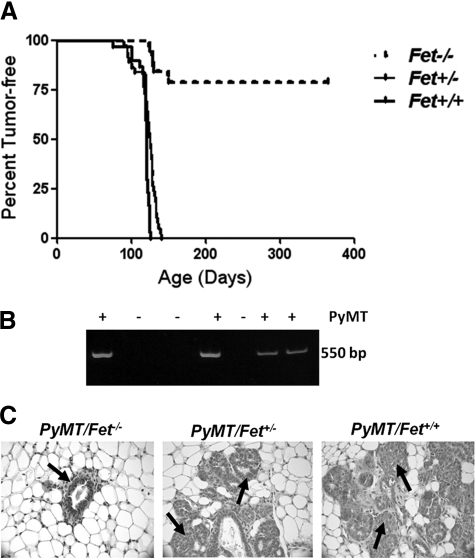
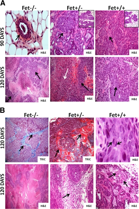
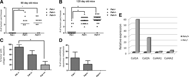
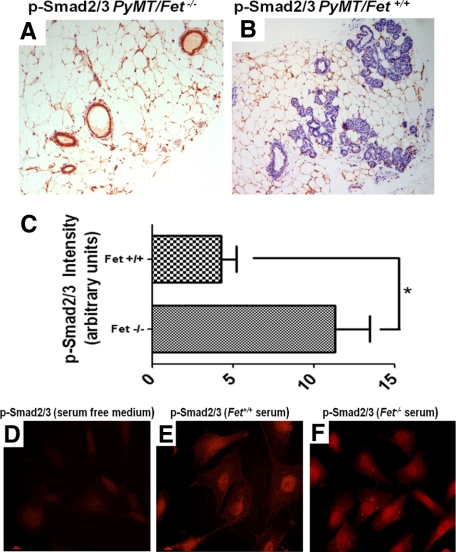
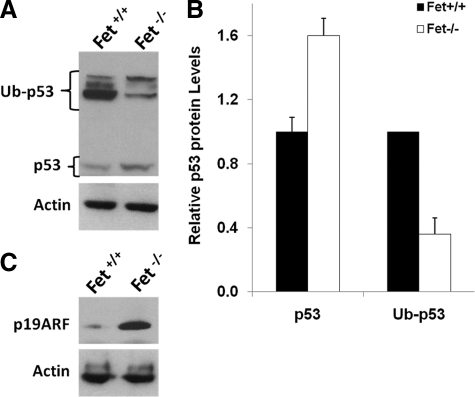
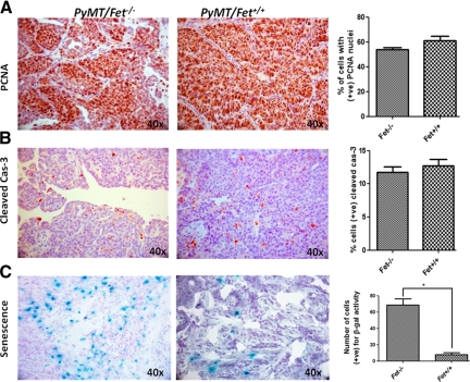
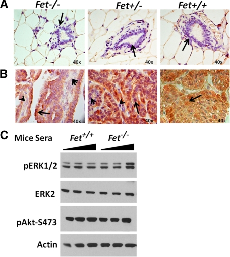
The present analyses were done to define the role of fetuin-A (Fet) in mammary tumorigenesis using the polyoma middle T antigen (PyMT) transgenic mouse model. We crossed Fet-null mice in the C57BL/6 background with PyMT mice in the same background and after a controlled breeding protocol obtained PyMT/Fet+/+, PyMT/Fet+/-, and PyMT/Fet-/- mice that were placed in control and experimental groups. Whereas the control group (PyMT/Fet+/+) formed mammary tumors 90 days after birth, tumor latency was prolonged in the PyMT/Fet-/- and PyMT/Fet+/- mice. The majority of the PyMT/Fet-/- mice were tumor-free at the end of the study, at approximately 40 weeks. The pathology of the mammary tumors in the Fet-null mice showed extensive fibrosis, necrosis, and squamous metaplasia. The preneoplastic mammary tissues of the PyMT/Fet-/- mice showed intense phopho-Smad2/3 staining relative to control tissues, indicating that transforming growth factor-β signaling is enhanced in these tissues in the absence of Fet. Likewise, p19ARF and p53 were highly expressed in tumor tissues of PyMT/Fet-/- mice relative to the controls in the absence of Fet. The phosphatidylinositol 3-kinase/Akt signaling pathway that we previously showed to be activated by Fet, on the other hand, was unaffected by the absence of Fet. The data indicate that Fet is a powerful modulator of breast tumorigenesis in this model system and has the potential to modulate breast cancer progression in humans.


Figures

Figure 1
Figure 1
Lack of fetuin-A decreases mammary tumor incidence and increases tumor latency in PyMT/Fet−/− relative to PyMT/Fet+/+ and PyMT/Fet+/− mice. A: Kaplan-Meier survival curve showing that all of the PyMT/Fet+/+ and PyMT/Fet+/− mice in the study succumbed to mammary tumors by approximately 180 days of age, whereas more than 60% of the PyMT/Fet−/− mice were tumor-free at the end of the experiment at approximately 380 days of age. B: A typical genotyping assay revealed the 550-bp band of the PyMT oncogene. C: Mammary tissues from 60-day-old mice. Note that the acinus of PyMT/Fet−/− looks normal (arrow), whereas the acini of PyMT/Fet+/− and PyMT/Fet+/+ exhibit typical changes of multifocal intraepithelial neoplasia containing multifocal areas of glandular differentiation (arrows).
Figure 2
Figure 2
Histopathology of mammary tissues of PyMT/Fet−/−, PyMT/Fet+/−, and PyMT/Fet+/+ at 90 and 120 days of age. A, top panel: Normal acinus (arrow) in a PyMT/Fet−/− mouse at 90 days of age (original magnification, ×40). The mammary tumor tissues of PyMT/Fet+/− and PyMT/Fet+/+ mice at 90 days of age clearly show tumor masses (arrows original magnification, ×20). There are also several hyperplastic acini and mammary intraepithelial neoplasm (insets). A, bottom panel: Tumors of PyMT/Fet−/− (original magnification, ×10), PyMT/Fet+/−, and PyMT/Fet+/+ (original magnification, ×20) mice show expansile tumor masses and extensive to moderate areas of necrosis (white arrows) interspersed with islands of neoplastic cells (black arrows). B, top panel: Mammary tumors show accumulation of collagen fibers particularly in tumors in PyMT/Fet−/− mice (stained blue with trichrome [TRIC], arrows). Tumor tissues of PyMT/Fet−/− and PyMT/Fet+/− also show squamous metaplasia and parakeratosis with concentric lamellar keratin consistent with keratin pearls (stained red with trichome; original magnification, ×20). Mitotic figures (arrows) for PyMT/Fet+/+ tumors of 3 to 5 per high-power field are shown (original magnification, ×40). B, bottom panel: tumor tissues in PyMT/Fet−/− mice show a medullary pattern (original magnification, ×10). This panel also shows mild to moderate mononuclear cell infiltration (arrows) in the tumor tissues of PyMT/Fet+/− and PyMT/Fet+/+ mice (original magnification, ×20).
Figure 3
Figure 3
Mammary tumor incidence at 90 and 120 days in the three PyMT genotypes. A: Number of palpable tumors/mouse in 90-day-old PyMT/Fet−/−, PyMT/Fet+/−, and PyMT/Fet+/+ mice (*P = 0.0002; **P = 0.0004). B: Number of palpable tumors/mouse in 120-day-old PyMT/Fet−/−, PyMT/Fet+/−, and PyMT/Fet+/+ mice. *P < 0.0001 C: Collagen deposition in the tumors of 120-day-old PyMT/Fet−/−, PyMT/Fet+/−, and PyMT/Fet+/+ mice as revealed by trichrome staining. *P < 0.05 D: Incidence of squamous metaplasia in the mammary tumors of 120-day-old PyMT/Fet−/− and PyMT/Fet+/− but not PyMT/Fet+/+ mice (mean ± SE, n = 10 mice per group; one-way analysis of variance). E: Real-time RT-PCR of collagen types 1 and 4 mRNA expression in the 120-day-old mammary tumors in PyMT/Fet−/− and PyMT/Fet+/+ animals.
Figure 4
Figure 4
TGF-β signaling determined by staining for P-Smad2/3 in mammary tissues of mice and human cells. A and B: Intensity of P-Smad2/3 staining in the mammary tissues of 60-day-old PyMT/Fet−/− (A) and PyMT/Fet+/+(B) mice (original magnification, ×10). C: Intensity of P-Smad2/3 staining represented as arbitrary units. *P = 0.008 D–F: TGF-β signaling in BT-549 breast tumor cells incubated in the absence of serum (D) and in the presence of serum from PyMT/Fet+/+ (E) and PyMT/Fet−/− (F) animals.
Figure 5
Figure 5
Fetuin-A-mediated inactivation of ARF-P19 and degradation of P53. Equal amounts of detergent-solubilized proteins of mammary tumors from the fetuin-A wild-type and null mice prepared as described in Materials and Methods were separated on a 4 to 12% SDS gel and transferred to nitrocellulose membranes, and the membranes were probed with antibodies to p53 (A) or p19ARF (C). Actin was used as a loading control. Note that the exposure of membranes probed with anti-p53 antibody was performed overnight at room temperature. Ub-p53, ubiquitinylated p53. B: Densitometric analysis of the p53 and Ub-p53 bands of whole tissue lysates. Bars represent means and SD of densitometric ratios relative to Fet+/+ from blots of two representative experiments.
Figure 6
Figure 6
Proliferation, apoptotic, and senescence markers in mammary tumors from PyMT/Fet−/− and PyMT/Fet+/+ animals. Mammary tumors (120 days old) from fetuin-A null and qjwild-type PyMT animals were sectioned and processed for immunohistochemical analysis as described in Materials and Methods. The slides were stained for PCNA (proliferation) (A) and cleaved caspase-3 (cas-3, apoptosis) (B). C: The slides were also stained for β-galactosidase activity that was quantified as number of cells positive for β-galactosidase (β-gal) activity per field (*P < 0.004). Original magnification, ×40. +ve, positive.
Figure 7
Figure 7
Expression of phospho-Akt-Ser-473 in the PyMT mammary tissues. A and B: Negligible expression of phospho-Akt-Ser-473 in normal mammary acini (A, arrows) (original magnification, ×40) and strong expression in mammary tumor sections (B, arrows) (original magnification, ×40). There were, however, some cells in the tumor sections of Fet−/− and Fet+/− animals in which phospho-Akt expression was weak (arrowheads). C: Breast carcinoma cells (BT-549) were incubated with graded doses (9.3–930 μg/ml) of serum proteins from PyMT/Fet+/+ and PyMT/Fet−/− mice for 30 minutes, followed by lysis of cells and resolution in SDS-polyacrylamide gel electrophoresis and transferred to immobile membrane. The membranes were probed with anti-phospho-Erk1/2 (pERK1/2), Erk2, phospho-Akt-473 (pAkt-S473), and actin. There was no marked difference in the level of activation of either Erk or Akt mediated by serum proteins from fetuin-A wild-type and null (minus fetuin-A) mice.

References

    1. Nie Z. Fetuin: its enigmatic property of growth promotion. Am J Physiol. 1992;263:C551–C562. - PubMed
    1. Jahnen-Dechent W, Schinke T, Trindl A, Muller-Esterl W, Sablitzky F, Kaiser S, Blessing M. Cloning and targeted deletion of the mouse fetuin gene. J Biol Chem. 1997;272:31496–31503. - PubMed
    1. Kundranda MN, Henderson M, Carter KJ, Gorden L, Binhazim A, Ray S, Baptiste T, Shokrani M, Leite-Browning ML, Jahnen-Dechent W, Matrisian LM, Ochieng J. The serum glycoprotein fetuin-A promotes Lewis lung carcinoma tumorigenesis via adhesive-dependent and adhesive-independent mechanisms. Cancer Res. 2005;65:499–506. - PubMed
    1. Swallow CJ, Partridge EA, Macmillan JC, Tajirian T, DiGuglielmo GM, Hay K, Szweras M, Jahnen-Dechent W, Wrana JL, Redston M, Gallinger S, Dennis JW. α2HS-glycoprotein, an antagonist of transforming growth factor β in vivo, inhibits intestinal tumor progression. Cancer Res. 2004;64:6402–6409. - PubMed
    1. Triffitt JT, Gebauer U, Ashton BA, Owen ME, Reynolds JJ. Origin of plasma α2-HS-glycoprotein and its accumulation in bone. Nature. 1976;262:226–227. - PubMed

Publication types

MeSH terms