

A molecular profile of focal segmental glomerulosclerosis from formalin-fixed, paraffin-embedded tissue
- PMID: 20847290
- PMCID: PMC2947265
- DOI: 10.2353/ajpath.2010.090746
A molecular profile of focal segmental glomerulosclerosis from formalin-fixed, paraffin-embedded tissue
Abstract
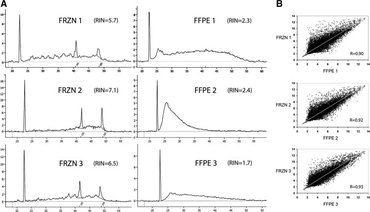
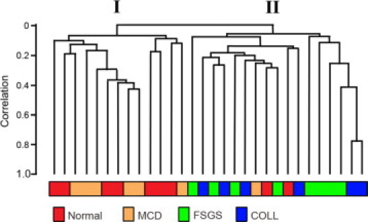
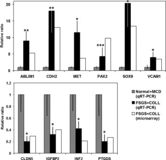
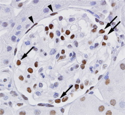
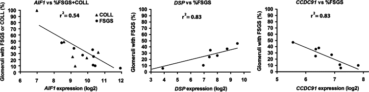
Focal segmental glomerulosclerosis (FSGS) is a common form of idiopathic nephrotic syndrome defined by the characteristic lesions of focal glomerular sclerosis and foot process effacement; however, its etiology and pathogenesis are unknown. We used mRNA isolated from laser-captured glomeruli from archived formalin-fixed, paraffin-embedded renal biopsies, until recently considered an unsuitable source of mRNA for microarray analysis, to investigate the glomerular gene expression profiles of patients with primary classic FSGS, collapsing FSGS (COLL), minimal change disease (MCD), and normal controls (Normal). Amplified mRNA was hybridized to an Affymetrix Human X3P array. Unsupervised (unbiased) hierarchical clustering revealed two distinct clusters delineating FSGS and COLL from Normal and MCD. Class comparison analysis of FSGS + COLL combined versus Normal + MCD revealed 316 significantly differentially regulated genes (134 up-regulated, 182 down-regulated). Among the differentially regulated genes were those known to be part of the slit diaphragm junctional complex and those previously described in the dysregulated podocyte phenotype. Analysis based on Gene Ontology categories revealed overrepresented biological processes of development, differentiation and morphogenesis, cell motility and migration, cytoskeleton organization, and signal transduction. Transcription factors associated with developmental processes were heavily overrepresented, indicating the importance of reactivation of developmental programs in the pathogenesis of FSGS. Our findings reveal novel insights into the molecular pathogenesis of glomerular injury and structural degeneration in FSGS.
Figures
References
-
- D'Agati VD, Fogo AB, Bruijn JA, Jennette JC. Pathologic classification of focal segmental glomerulosclerosis: a working proposal. Am J Kidney Dis. 2004;43:368–382. - PubMed
-
- Korbet SM. Primary focal segmental glomerulosclerosis. J Am Soc Nephrol. 1998;9:1333–1340. - PubMed
-
- Stokes MB, Valeri AM, Markowitz GS, D'Agati VD. Cellular focal segmental glomerulosclerosis: clinical and pathologic features. Kidney Int. 2006;70:1783–1792. - PubMed
-
- Thomas DB, Franceschini N, Hogan SL, Ten Holder S, Jennette CE, Falk RJ, Jennette JC. Clinical and pathologic characteristics of focal segmental glomerulosclerosis pathologic variants. Kidney Int. 2006;69:920–926. - PubMed
-
- D'Agati VD. The spectrum of focal segmental glomerulosclerosis: new insights. Curr Opin Nephrol Hypertens. 2008;17:271–281. - PubMed
Publication types
MeSH terms
Substances
Grants and funding
LinkOut - more resources
Full Text Sources
Other Literature Sources
