

Increased blood-brain barrier permeability on perfusion CT might predict malignant middle cerebral artery infarction
- PMID: 20847316
- PMCID: PMC3412880
- DOI: 10.1161/STROKEAHA.110.591362
Increased blood-brain barrier permeability on perfusion CT might predict malignant middle cerebral artery infarction
Abstract
Background and purpose: Perfusion CT has been used to assess the extent of blood-brain barrier breakdown. The purpose of this study was to determine the predictive value of blood-brain barrier permeability measured using perfusion CT for development of malignant middle cerebral artery infarction requiring hemicraniectomy (HC).
Methods: We retrospectively identified patients from our stroke registry who had middle cerebral artery infarction and were evaluated with admission perfusion CT. Blood-brain barrier permeability and cerebral blood volume maps were generated and infarct volumes calculated. Clinical and radiographic characteristics were compared between those who underwent HC versus those who did not undergo HC.
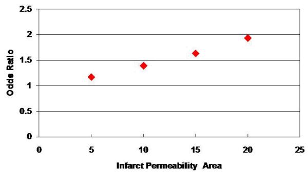
Results: One hundred twenty-two patients (12 HC, 110 no HC) were identified. Twelve patients who underwent HC had developed edema, midline shift, or infarct expansion. Infarct permeability area, infarct cerebral blood volume area, and infarct volumes were significantly different (P < 0.018, P < 0.0211, P < 0.0001, P < 0.0014) between HC and no HC groups. Age (P = 0.03) and admission National Institutes of Health Stroke Scale (P = 0.0029) were found to be independent predictors for HC. Using logistic regression modeling, there was an association between increased infarct permeability area and HC. The OR for HC based on a 5-, 10-, 15-, or 20-cm² increase in infarct permeability area were 1.179, 1.390, 1.638, or 1.932, respectively (95% CI, 1.035 to 1.343, 1.071 to 1.804, 1.108 to 2.423, 1.146 to 3.255, respectively).
Conclusions: Increased infarct permeability area is associated with an increased likelihood for undergoing HC. Because early HC for malignant middle cerebral artery infarction has been associated with better outcomes, the infarct permeability area on admission perfusion CT might be a useful tool to predict malignant middle cerebral artery infarction and need for HC.
Figures

References
-
- Berrouschot J, Sterker M, Bettin S, Köster J, Schneider D. Mortality of space-occupying (‘malignant’) middle cerebral artery infarction under conservative intensive care. Intensive Care Med. 1998;24:620–3. - PubMed
-
- Hacke W, Schwab S, Horn M, Spranger M, De Georgia M, von Kummer R. ‘Malignant’ middle cerebral artery territory infarction: clinical course and prognostic signs. Arch Neurol. 1996;53:309–15. - PubMed
-
- Rieke K, Schwab S, Krieger D, von Kummer R, Aschoff A, Schuchardt V, Hacke W. Decompressive surgery in space-occupying hemispheric infarction: results of an open, prospective trial. Crit Care Med. 1995;23:1576–87. - PubMed
-
- Schwab S, Rieke K, Aschoff A, Albert F, von Kummer R, Hacke W. Hemicraniotomy in space-occupying hemispheric infarction:Useful early intervention or desparate activism? Cerebrovasc Dis. 1996;6:325–329.
-
- Schwab S, Steiner T, Aschoff A, Schwarz S, Steiner HH, Jansen O, Hacke W. Early hemicraniectomy in patients with complete middle cerebral artery infarction. Stroke. 1998;29:1888–93. - PubMed
