

Accessory Breast Carcinoma
- PMID: 20847887
- PMCID: PMC2931069
- DOI: 10.1159/000210638
Accessory Breast Carcinoma
Abstract
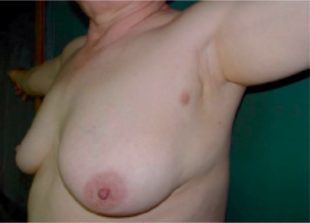
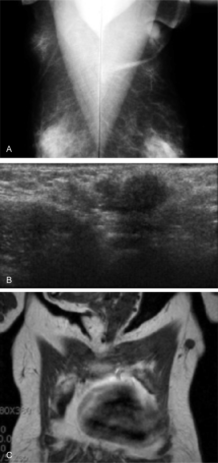
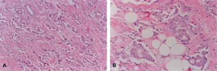
SUMMARY: BACKGROUND: Ectopic breast tissue usually develops along the mammary ridges, and the incidence has been reported to be 2-6% of the general population. Occurrence of primary carcinoma in ectopic breast tissue is rare. CASE REPORT: We report the case of 59-year-old woman with accessory breast carcinoma in her left axilla. CONCLUSION: Because an accessory areola or nipple is often missing and awareness of physicians and patients about these unsuspicious masses is lacking, clinical diagnosis of accessory breast carcinoma is frequently delayed. Therefore, a mass along the 'milk line' should be examined carefully, and any suspicious lesions should be evaluated.
Hintergrund: Ektopisches Brustgewebe entwickelt sich gewöhnlich entlang der Milchleiste mit einer Inzidenz von 2–6% in der allgemeinen Bevölkerung. Das Auftreten primärer Karzinome in ektopischem Brustgewebe ist selten.
Fallbericht: Wir berichten von einer 59-jährigen Patientin mit einem Mammakarzinom in einer akzesso-rischen Brustdrüse in der linken Axilla.
Schlussfolgerung: Da akzessorische Warzenhöfe oder Brustwarzen zumeist nicht vorhanden sind und sowohl Ärzte als auch Patienten die meist unverdächtigen Knoten oft nicht zur Kenntnis nehmen, ist die klinische Diagnosestellung bei Mammakarzinomen akzessorischer Brustdrüsen regelmäßig verzögert. Ein Knoten entlang der Milchleiste sollte daher eingehend untersucht und verdächtige Läsionen genauer geprüft werden.
Figures
References
-
- Marshall MB, Moynihan JJ, Frost A, Evans SR. Ectopic breast cancer: case report and literature review. Surg Oncol. 1994;3:295–304. - PubMed
-
- Evans DM, Guyton DP. Carcinoma of the axillary breast. J Surg Oncol. 1995;59:190–195. - PubMed
-
- Yerra L, Karnad AB, Votaw ML. Primary breast cancer in aberrant breast tissue in the axilla. South Med J. 1997;90:661–662. - PubMed
-
- Camisa C. Accessory breast on the posterior thigh of a man. J Am Acad Dermatol. 1980;3:467–469. - PubMed
-
- Copeland MM, Geschickter CF. Symposium on diagnosis and treatment of premalignant conditions. Surg Clins N Am. 1950;30:1717–1741. - PubMed
Publication types
LinkOut - more resources
Full Text Sources
