. 2010 Nov;28(11):2027-40.
doi: 10.1002/stem.525.

Convergent genesis of an adult neural crest-like dermal stem cell from distinct developmental origins

Affiliations

Convergent genesis of an adult neural crest-like dermal stem cell from distinct developmental origins

Hiroyuki Jinno et al. Stem Cells. 2010 Nov.

Abstract

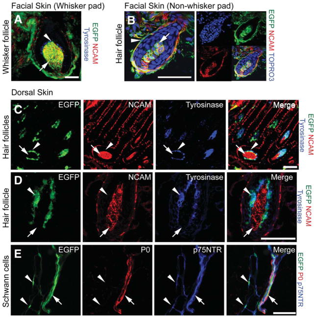
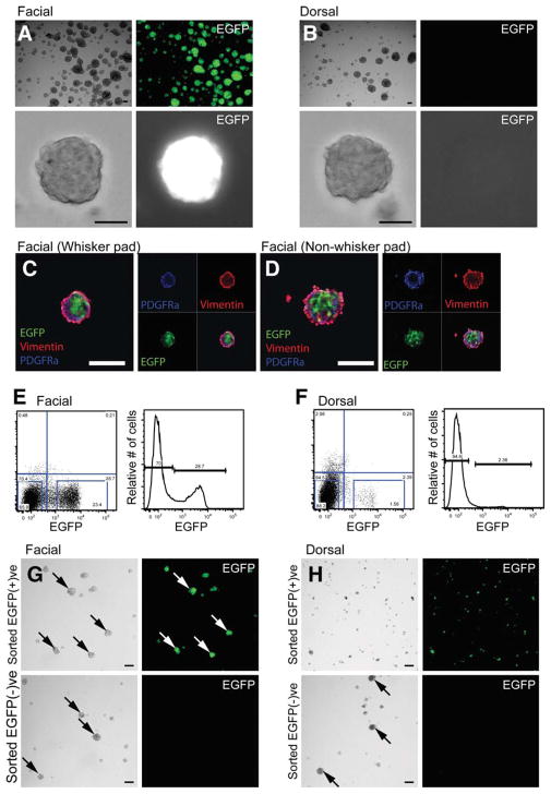
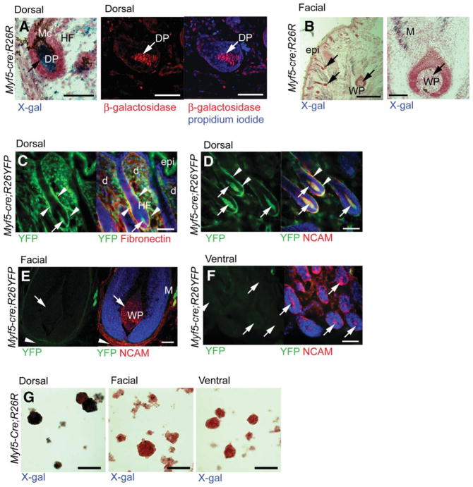
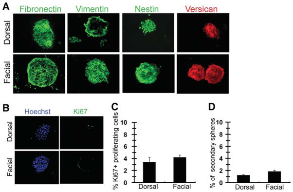
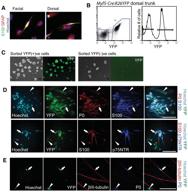
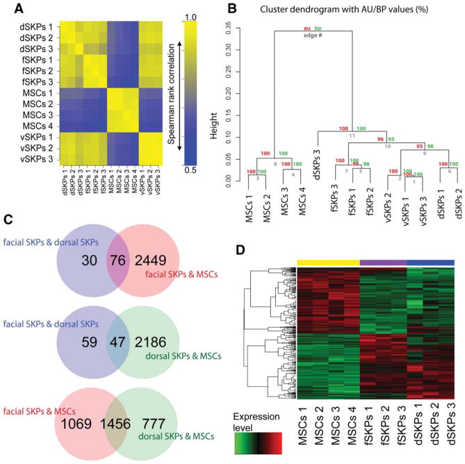
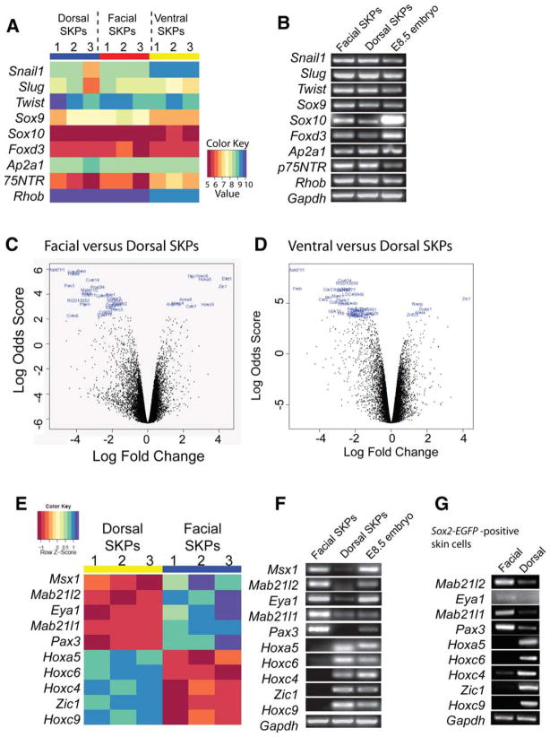
Skin-derived precursors (SKPs) are multipotent dermal stem cells that reside within a hair follicle niche and that share properties with embryonic neural crest precursors. Here, we have asked whether SKPs and their endogenous dermal precursors originate from the neural crest or whether, like the dermis itself, they originate from multiple developmental origins. To do this, we used two different mouse Cre lines that allow us to perform lineage tracing: Wnt1-cre, which targets cells deriving from the neural crest, and Myf5-cre, which targets cells of a somite origin. By crossing these Cre lines to reporter mice, we show that the endogenous follicle-associated dermal precursors in the face derive from the neural crest, and those in the dorsal trunk derive from the somites, as do the SKPs they generate. Despite these different developmental origins, SKPs from these two locations are functionally similar, even with regard to their ability to differentiate into Schwann cells, a cell type only thought to be generated from the neural crest. Analysis of global gene expression using microarrays confirmed that facial and dorsal SKPs exhibit a very high degree of similarity, and that they are also very similar to SKPs derived from ventral dermis, which has a lateral plate origin. However, these developmentally distinct SKPs also retain differential expression of a small number of genes that reflect their developmental origins. Thus, an adult neural crest-like dermal precursor can be generated from a non-neural crest origin, a finding with broad implications for the many neuroendocrine cells in the body.


Conflict of interest statement

Disclosure of Potential Conflicts of Interest

The authors indicate no potential conflicts of interest.

Figures

Figure 1
Figure 1
In facial but not dorsal trunk skin, follicle dermal papilla (DP) and DS derive from the neural crest. (A, B): Immunocytochemistry of facial skin sections from neonatal Wnt1-cre;Z/EG mice. (A): Whisker follicle section immunostained for EGFP (green), the DP marker NCAM (red), and the melanoblast/melanocyte marker tyrosinase (blue). The arrow denotes the whisker follicle DP, and the arrowhead the overlying melanocytes. (B): A section through a hair follicle in facial skin outside of the whisker pad immunostained for EGFP (green) and the DP marker NCAM (red). Nuclei were stained with the dye TOPRO3 (blue). The arrow denotes the hair follicle DP, and the arrowhead the dermal sheath. (C, D): Dorsal trunk skin sections from neonatal Wnt1-cre;Z/EG mice, immunostained for EGFP (green), the DP marker NCAM (red), and the melanoblast/melanocyte marker tyrosinase (blue). Arrows denote the DP of dorsal hair follicles and arrowheads the overlying melanocytes. (E): Dorsal skin from neonatal Wnt1-cre:Z/EG mice, immunostained for EGFP (green) and the Schwann cell markers P0 peripheral myelin protein (red) and p75NTR (blue). Arrows denote a nerve containing myelinating Schwann cells positive for all three markers, whereas arrowheads indicate EGFP-positive, p75NTR-positive nonmyelinating Schwann cells. Scale bar = 50 μm (for all panels). Abbreviations: EGFP, enhanced green fluorescent protein; NCAM, neural cell adhesion molecule.
Figure 2
Figure 2
In facial but not dorsal trunk skin, skin-derived precursors (SKPs) derive from neural crest. (A, B): Primary SKP spheres generated from neonatal Wnt1-cre;Z/EG facial (A) and dorsal trunk (B) skin. In the bottom panels, single spheres are shown at high magnification. Scale bar = 50 μm. (C, D): Primary facial SKP spheres from whisker pad skin and from facial skin outside of the whisker pad, immunostained for EGFP (green), and the SKP markers vimentin (red) and PDGFRα (blue). Scale bar = 100 μm. (E, F): Flow cytometry plots of neonatal Wnt1-cre;Z/EG facial (E) and dorsal trunk (F) skin, sorted on the basis of EGFP expression. Left panels show the primary data, with the bottom blue boxes indicating the cells that were collected for culture. Numbers represent the percentage of cells in each quadrant of the plot. Right panels show the data plotted as relative numbers of cells expressing no or high relative levels of EGFP. The percentage of cells in each of those two groups is indicated. (G, H): Skin cells sorted as in (E, F) were cultured in SKPs medium for 1–2 weeks, passaged, and photographed 1 week later. Arrows indicate secondary SKP spheres. For facial skin (G), the EGFP-positive cell fraction contained most of the SKP-forming activity, although some SKP spheres were generated from the EGFP-negative population. For dorsal trunk skin (H), EGFP-positive cells did not generate SKP spheres when passaged, but remained as single cells or small clumps of cells, and virtually all of the SKP-forming activity was present within the EGFP-negative cell fraction. Scale bar = 100 μm. Abbreviations: EGFP, enhanced green fluorescent protein; PDGFRα, platelet derived growth factor receptor α.
Figure 3
Figure 3
In dorsal trunk but not facial or ventral trunk skin, hair follicle DP/dermal sheath (DS) and Skin-derived precursors (SKPs) derive from the somites. (A, B): Dorsal trunk (A) and facial (B) skin sections from neonatal Myf5-cre;R26R mice, stained with X-gal ([A], left panel and [B], blue) or immunostained for β-galactosidase ([A], middle and right panels, red) to detect β-galactosidase. Tissue was counterstained with nuclear fast red (red in [A], left panel and [B]) or propidium iodide (blue in [A], right panel) to show morphology. Arrows denote hair and whisker follicle DP. Scale bar = 50 μm (A), 500 μm ([B], left) and 100 μm ([B], right). (C–F): Dorsal trunk (C, D), facial (E), and ventral trunk (F) skin sections from neonatal Myf5-cre;R26YFP mice, immunostained for EYFP (green), fibronectin ([C], red) or NCAM ([D–F], red). Tissues were counterstained with the nuclear dye TOPRO3 (blue) to show morphology. Arrows denote follicle DP, which are EYFP-positive in dorsal trunk skin (C, D), but not in facial (E) or ventral trunk (F) skin. Arrowheads denote EYFP-positive DS cells in dorsal trunk skin (C, D). Note that muscle cells (M) in facial skin (E) are EYFP-positive. Scale bar = 50 μm. (G): Secondary SKP spheres generated from neonatal Myf5-cre;R26R dorsal trunk skin (left panel), facial skin (middle panel), and ventral trunk skin (right panel), stained with X-gal to detect β-galactosidase (blue) and counterstained with nuclear fast red (red). Scale bar = 200 μm. Abbreviations: d, interfollicular dermis; DP, dermal papilla; epi, epidermis; HF, hair follicle; M, muscle; Mc, melanocytes; NCAM, neural cell adhesion molecule; WP, whisker papilla; YFP, yellow fluorescent protein.
Figure 4
Figure 4
Facial and dorsal trunk skin-derived precursors (SKPs) display similar properties. (A): Secondary SKP spheres generated from neonatal mouse dorsal trunk skin and facial skin, immunostained for fibronectin, vimentin, nestin, and versican. (B): Primary SKP spheres isolated from neonatal wild-type dorsal trunk skin and facial skin, immunostained for Ki67 (green), and counterstained with Hoechst to show cell nuclei (blue). (C): Quantification of Ki67-positive cells from experiments similar to that shown in (B). n = 3 independent experiments. (D): Quantification of the percentage of cells that form a new sphere from primary SKPs plated at clonal density (2,500 cells/ml) in methylcellulose cultures. n = 3 independent experiments.
Figure 5
Figure 5
Somite-derived skin-derived precursors (SKPs) generate Schwann cells. (A): Neonatal mouse dorsal trunk and facial skin cells cultured as SKPs, differentiated under gliogenic conditions for 2–3 weeks, and immunostained for the Schwann cell markers S100β (green) and GFAP (red), and counterstained with Hoechst (blue). (B): Flow cytometry plots of Myf5-cre;R26YFP dorsal trunk skin cells, sorted for relative levels of EYFP expression. The left panel shows the primary data, and the blue boxes show the cell fractions that were cultured as EYFP-positive versus negative cells. The right panel shows the same data plotted as relative cell numbers versus EYFP expression. The percentage of cells in each fraction is indicated. (C): Cells sorted as in (B) and cultured for 7 days in SKPs conditions. The left two panels show the SKP spheres derived from the EYFP-positive fraction and the right two panels show the EYFP-negative fraction, with the left panels of each pair showing phase illumination and the right the same fields with fluorescence illumination. (D): Myf5-cre;R26YFP-positive dorsal trunk SKPs were differentiated under gliogenic conditions for 2–3 weeks, and immunostained for EYFP (green), and the Schwann cell markers P0 (red, top panels) S100β (blue, top panels; red bottom panels) and p75NTR (blue, bottom panels). Arrows in top panels indicate EYFP-positive, P0-positive, S100β-positive spindle-shaped Schwann cells and in the bottom panels indicate EYFP-positive, S100β-positive, p75NTR-positive Schwann cells. Arrowheads indicate EYFP-positive, flat cells that are negative for the Schwann cell markers. Cells were counterstained with Hoechst (turquoise). Scale bar = 100 μm. (E): Differentiated Myf5-cre;R26YFP-positive dorsal trunk SKPs were cocultured with axons of sympathetic neurons in compartmented cultures for 8 days, and immunostained for EYFP (green), the Schwann cell marker P0 (blue), and the axonal marker βIII-tubulin (red). Arrows indicate a representative EYFP-positive, P0-positive, spindle-shaped Schwann cell associated with a βIII-tubulin-positive axon. Arrowheads indicate an EYFP-positive, flat cell that is not associated with an axon and that is negative for Schwann cell markers. Cells were counterstained with Hoechst (turquoise). Scale bar = 100 μm. Abbreviations: GFAP, glial fibrillary acidic protein; YFP, yellow fluorescent protein.
Figure 6
Figure 6
Microarray analysis of adult dorsal trunk, ventral trunk, and facial SKPs versus MSCs. Microarray analysis was performed to compare gene expression patterns among adult rat dorsal trunk, facial, and ventral trunk SKPs. Adult rat bone marrow MSCs were used as a comparator. Three independent isolates of secondary passage SKPs from dorsal trunk skin (dSKPs 1–3), facial skin (fSKPs 1–3), ventral trunk skin (fSKPs 1–3), and four isolates of MSCs (MSCs 1–4) were compared. (A): Spearman rank correlation matrix computed for the microarray experiments based on the 3,182 probesets showing the most variation across the experiments, as visualized by color-coding, with yellow representing the most highly correlated samples, and blue the least correlated. Note that the dorsal trunk, facial, and ventral trunk SKP samples are highly correlated with each other, whereas they show less correlation with the MSCs. (B): Microarray datasets from all four sets of samples were clustered using hierarchical clustering with correlation distance and average linkage. The significance of the hierarchical clustering result was assessed using AU and BP resampling implemented in the R package pvclust. (C): Venn diagrams of pairwise comparisons between facial SKPs, dorsal trunk SKPs, and MSCs to identify genes differentially expressed between each pair of samples using an analysis similar to one-way ANOVA implemented in the LIMMA bioconductor package. The Venn diagrams show significantly differentially expressed genes (p < .05, Benjamini) that are in common among the pairwise comparisons, revealing that facial SKPs and dorsal SKPs are more similar to each other than either of them are to MSCs. (D): Three-way comparison was conducted across the groups to identify genes that show evidence of differential expression (analysis similar to one-way analysis of variance). Expression profiles of 2,603 genes, identified as differentially expressed (p < .05, Benjamini), are plotted as a heatmap. Abbreviations: AU, approximately unbiased; BP, bootstrap probability; MSC, mesenchymal stromal cell; SKP, skin-derived precursor.
Figure 7
Figure 7
SKPs of all developmental origins express neural crest signature genes but retain a lineage history at the gene expression level. (A): Microarray expression levels of genes expressed in embryonic neural crest precursors in adult rat facial, dorsal trunk, and ventral trunk SKPs, plotted as a heatmap. Red indicates the lowest relative levels of expression and dark blue the highest, as defined by the color key. (B): Reverse transcription polymerase chain reactions (RT-PCRs) for the same genes shown in (A), in total RNA isolated from neonatal murine dorsal trunk and facial secondary SKP spheres. Total RNA from E8.5 murine embryos was used as a positive control. (C, D): Pairwise differential expression analysis was conducted between (C) facial and dorsal trunk SKPs and (D) ventral trunk and dorsal trunk SKPs from adult rats using the LIMMA bioconductor package. The 35 genes showing most significant differential expression between the two populations on the volcano plots are shown and are listed in Supporting Information Tables 1 and 2, after multiple testing correction. The positive log fold changes indicate genes that are expressed at higher levels in dorsal SKPs (C, D), and the negative log fold changes those that are expressed at decreased levels in dorsal SKPs relative to facial SKPs (C) or ventral trunk SKPs (D). (E): Microarray expression levels of transcription factors that were identified as being among the most differentially expressed in the analysis in (C), plotted as a heatmap. (F): RT-PCRs for the genes highlighted in (E) in total RNA from neonatal murine dorsal and facial secondary SKP spheres, highlighting the differential expression. Total RNA from E8.5 murine embryos was used as a positive control. (G): RT-PCRs for the genes highlighted in (F) in total RNA from uncultured, purified EGFP-positive cells from neonatal Sox2-EGFP mouse dorsal trunk and facial skin, highlighting differential expression in uncultured dermal precursors. Abbreviations: EGFP, enhanced green fluorescent protein; SKP, skin-derived precursor.

Similar articles

Cited by

References

    1. Toma JG, Akhavan M, Fernandes KJ, et al. Isolation of multipotent adult stem cells from the dermis of mammalian skin. Nat Cell Biol. 2001;3:778–784. - PubMed
    1. Lavoie JF, Biernaskie JA, Chen Y, et al. Skin-derived precursors differentiate into skeletogenic cell types and contribute to bone repair. Stem Cells Dev. 2009;18:893–906. - PubMed
    1. McKenzie IA, Biernaskie J, Toma JG, et al. Skin-derived precursors generate myelinating Schwann cells for the injured and dysmyelinated nervous system. J Neurosci. 2006;26:6651–6660. - PMC - PubMed
    1. Biernaskie JA, McKenzie IA, Toma JG, et al. Isolation of skin-derived precursors (SKPs) and differentiation and enrichment of their Schwann cell progeny. Nat Protoc. 2006;1:2803–2812. - PubMed
    1. Biernaskie J, Sparling JS, Liu J, et al. Skin-derived precursors generate myelinating Schwann cells that promote remyelination and functional recovery after contusion spinal cord injury. J Neurosci. 2007;27:9545–9559. - PMC - PubMed

Publication types