. 2010 Dec;101(12):2637-43.
doi: 10.1111/j.1349-7006.2010.01723.x. Epub 2010 Sep 17.

Incorporation of multicellular spheroids into 3-D polymeric scaffolds provides an improved tumor model for screening anticancer drugs

Affiliations

Incorporation of multicellular spheroids into 3-D polymeric scaffolds provides an improved tumor model for screening anticancer drugs

Won Jin Ho et al. Cancer Sci. 2010 Dec.

Abstract

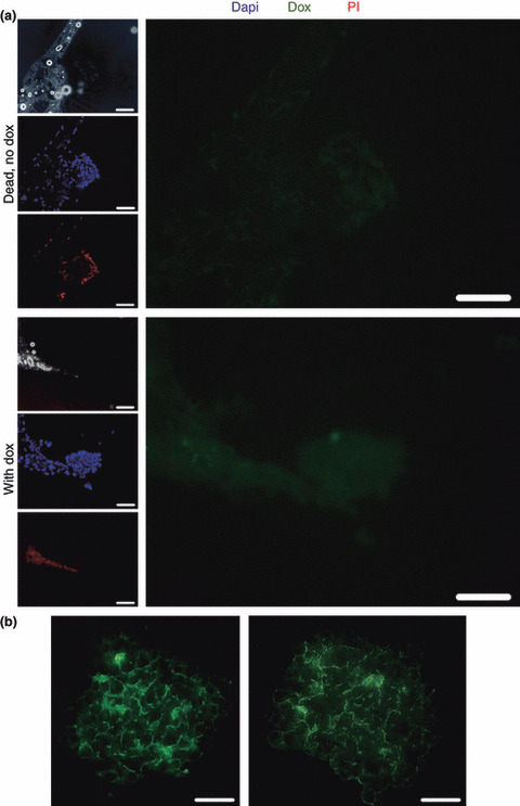
Development of cancer therapeutics requires a thorough evaluation of drug efficacy in vitro before animal testing and subsequent clinical trials. Three-dimensional (3-D) in vitro models have therefore been investigated for drug screening. In this study, we have developed a novel in vitro model in which multicellular aggregates, or spheroids, were incorporated into 3-D porous scaffolds. Drug resistance assays showed that spheroid-seeded scaffolds have much higher drug resistance than monolayer cultures, spheroids on flat substrates, or scaffolds seeded with dispersed cells. Furthermore, spheroid-seeded scaffolds demonstrated higher lactate production leading to acidosis, and higher expression of angiogenic factors. These data suggest that the spheroid-seeded 3-D scaffolds might serve as a useful in vitro system for screening cancer therapeutics.


Figures

Figure 1
Figure 1
Establishing spheroid‐seeded scaffolds. (a) Previously established in vitro tumor models include monolayer cultures, spheroids and scaffolds with cells. A novel tumor model is developed by incorporating spheroids, or 3‐D cellular aggregates, into 3‐D scaffolds. (b) Microscopic image of a 2‐day‐old spheroid of 2000 cells confirmed the spherical morphology. (c) Spheroid diameter gradually decreased over the duration of the culture (n = 10). Mean diameters (±SEM) are plotted as a function of time. (d) The viability of cells within spheroids cultured for 4 days was observed using a live stain (calcein‐AM). The control represents a dead spheroid that had been incubated in 70% ethanol for 30 min. (e) Images of poly(lactic‐co‐glycolic acid) (PLGA) scaffolds showed that spheroids were successfully incorporated into the pores of the scaffold. Scale bar, 100 μm.
Figure 2
Figure 2
Resistance to anticancer drugs. Four different systems, 2‐D at 20 000 cells/cm2 (▪), spheroids in 2‐D (•; only for doxorubicin), 3‐D scaffolds with monolayer‐cultured cells (MS, ), and 3‐D scaffolds with spheroid‐cultured cells (SS, ) were treated with (a) doxorubicin and (b) irinotecan and cell death was quantified. Mean percentage of cell deaths (±SEM) plotted as a function of drug concentration (n = 4).
Figure 3
Figure 3
Effects of reduced drug‐to‐cell ratio and polymer drug sequestering on drug resistance. To assess the significance of drug‐to‐cell ratio on the observed drug resistance, four different cell densities (10 k/cm2, 20 k/cm2, 40 k/cm2 and 60 k/cm2) were subjected to varying concentrations of either doxorubicin or irinotecan, and cell viability was quantified. Mean percentage of cell deaths (±SEM, n = 3–4) were plotted as a function of concentration of (a) doxorubicin or (b) irinotecan. The amount of doxorubicin sequestered onto poly(lactic‐co‐glycolic acid) (PLGA) scaffolds was also quantified. Bar graph (c) represents μg of doxorubicin sequestered (mean + SEM, n = 3). ***P < 0.001.
Figure 4
Figure 4
Assessing drug penetration. (a) Visualization of doxorubicin (dox) penetration into spheroids within the scaffold was based on doxorubicin’s intrinsic fluorescence (white: negative bright field; blue (middle left panel): DAPI; green (right panel): doxorubicin; red (bottom left panel): propidium iodide [PI]; scale bar, 100 μm). Controls include SS incubated in culture medium without doxorubicin and blank collagen‐coated scaffolds incubated in propidium iodide only (not shown). (b) The presence of intercellular spaces within the spheroids were visualized by incubating SS in PBS containing fluorescein isothiocyanate‐conjugated 150 kD dextran (scale bar, 50 μm).
Figure 5
Figure 5
Hypoxia‐associated characteristics of spheroid‐seeded scaffolds. (a) The amount of lactate produced by each tumor model was quantified using a lactate assay kit. All data (mean + SEM, n = 4–8) are normalized to 2‐D values. ***P < 0.001. (b) Degree of acidosis in SS was also evaluated by measuring the intracellular pH. Exterior represents the 50‐μm‐wide perimeter of spheroids within the scaffolds, while interior is defined as the inner core of the spheroids. Mean pH values (±SEM) from the two regions are plotted (n = 4). *P < 0.05. (c) The relative production of angiogenic factors, VEGF and bFGF were also quantified. All data (mean + SEM, n = 3–4) are normalized to their respective 2‐D values. *P < 0.05. **P < 0.01. ***P < 0.001 (all compared with 2‐D).

References

    1. Griffith L, Swartz M. Capturing complex 3D tissue physiology in vitro. Nat Rev Mol Cell Biol 2006; 7: 211–24. - PubMed
    1. Newman MJ, Rodarte JC, Benbatoul KD et al. Discovery and characterization of OC144‐093, a novel inhibitor of P‐glycoprotein‐mediated multidrug resistance. Cancer Res 2000; 60: 2964–72. - PubMed
    1. Torrance CJ, Agrawal V, Vogelstein B, Kinzler KW. Use of isogenic human cancer cells for high‐throughput screening and drug discovery. Nat Biotechnol 2001; 19: 940–5. - PubMed
    1. Holtfreter J. A study of the mechanics of gastrulation. J Exp Zool 1944; 95: 171–212.
    1. Moscona A. Cell suspensions from organ rudiments of chick embryos. Exp Cell Res 1952; 3: 535–9.

MeSH terms