. 2011 Jan 20;488(2):112-7.
doi: 10.1016/j.neulet.2010.09.018. Epub 2010 Sep 19.

IGF-I stimulates Rab7-RILP interaction during neuronal autophagy

Affiliations

IGF-I stimulates Rab7-RILP interaction during neuronal autophagy

Mona Bains et al. Neurosci Lett. .

Abstract

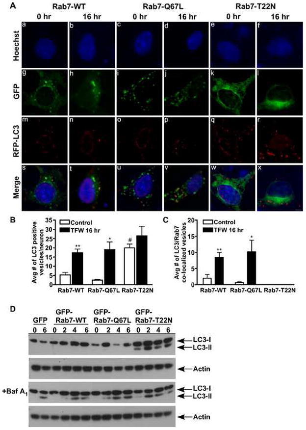
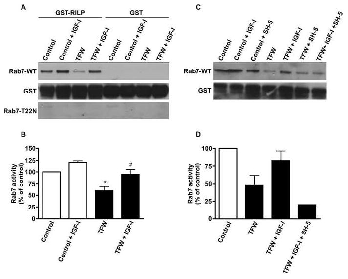
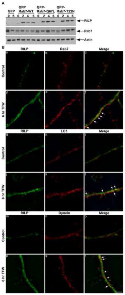
Restoration of autophagy represents a potential therapeutic target for neurodegenerative disorders, but factors that regulate autophagic flux are largely unknown. When deprived of trophic factors, cultured Purkinje neurons die by an autophagy associated cell death mechanism. The accumulation of autophagic vesicles and cell death of Purkinje neurons is inhibited by insulin-like growth factor, by a mechanism that enhances autophagic vesicle turnover. In this report, we identify Rab7 as an IGF-I regulated target during neuronal autophagy. Purkinje neurons transfected with EGFP-Rab7-WT and constitutively active EGFP-Rab7-Q67L contained few RFP-LC3 positive autophagosomes and little co-localization with GFP-Rab7 under control conditions. Upon induction of autophagy, RFP-LC3 positive autophagosomes increased and co-localized with GFP-Rab7. Conversely, expression of the dominant negative mutant EGFP-Rab7-T22N increased the accumulation of autophagosomes under control conditions, which accumulated even further during trophic factor withdrawal. There was no vesicular co-localization between Rab7-T22N and RFP-LC3 under control or trophic factor withdrawal conditions. During prolonged trophic factor withdrawal, a condition that leads to the accumulation of autophagic vesicles and cell death, Rab7 activity decreased significantly. IGF-I, added at the time of trophic factor withdrawal, prevented the deactivation of Rab7 and increased the interaction of Rab7 with its interacting protein (RILP), restoring autophagic flux. These results provide a novel mechanism by which IGF-I regulates autophagic flux during neuronal stress.


Figures

Fig. 1
Fig. 1
Rab7 regulates autophagic flux in Purkinje neurons. (A) Cerebellar cultures were transfected with GFP or Rab7-WT, Rab7-Q67L, or Rab7-T22N tagged to GFP. Neurons were infected with RFP-LC3, incubated in control or TFW media for 16 hr and subjected to live-cell imaging. Scale bar represents 5 μm. The total number of autophagosomes (B) and Rab7/LC3 co-localized vesicles (C) were counted in 10 Purkinje neurons per treatment condition and averaged for three separate experiments. (D) Rab7 transfected neurons were treated with control or TFW media in the absence or presence of bafilomycin A1 (100 nM) for 0, 2, 4 and 6 hr and an immunoblot analysis was performed against LC3. Actin served as a loading control. Figure 1D is a representative immunoblot of three independent experiments. Data represent mean ± SEM of three independent experiments. (B) **p<0.01 compared to Rab7-WT control, *p<0.05 compared to Rab7-Q67L control, #p<0.05 compared to Rab7-WT and Rab7-Q67L control (C) **p<0.01 compared to Rab7-WT control, *p<0.05 compared to Rab7-Q67L control. One-way ANOVA followed by Tukey’s post hoc test.
Fig. 2
Fig. 2
Rab7 activity during TFW-induced autophagy is regulated by IGF-I. (A) and (C) Cerebellar cultures were transfected with GFP or GFP-tagged Rab7-WT or Rab7-T22N and subjected to 16 hr of TFW in the absence or presence of IGF-I (200 ng/ml) or IGF-I plus the Akt inhibitor, SH-5 (1.0 μm). GST or GST-RILP were added to the cell lysates to precipitate active GTP-bound Rab7. Total GST served as a loading control. (B,D) Quantification of results shown in (A and C). The amount of Rab7 detected in the GST-RILP pull-down was normalized to GST and expressed as Rab7 activity, percent of control. Data represent mean ± SEM of three independent experiments. (B) *p<0.05 compared to control, #p<0.05 compared to TFW. One-way ANOVA followed by Tukey’s post hoc test.
Fig. 3
Fig. 3
RILP expression levels and localization during TFW-induced autophagy. (A) Cerebellar cultures were transfected with GFP or Rab7-WT, Rab7-Q67L or Rab7-T22N tagged to GFP. Neurons were treated in the absence or presence of trophic factors for 0, 2, 4 and 6 hr. Immunoblot analysis was performed using a polyclonal antibody to RILP and Rab7. Actin served as a loading control. (B) Cerebellar cultures were maintained in control or TFW medium for 6 hr, fixed and stained simultaneously with antibodies against RILP (green) and Rab7 (red), RILP (green) and LC3 (red) or RILP (green) and dynein (red). Shown are representative images of the immunofluorescence findings. Scale bar represents 5 μm.

References

    1. Anglade P, Vyas S, Javoy-Agid F, Herrero MT, Michel PP, Marquez J, Mouatt-Prigent A, Ruberg M, Hirsch EC, Agid Y. Apoptosis and autophagy in nigral neurons of patients with Parkinson’s disease. Histol Histopathol. 1997;12:25–31. - PubMed
    1. Bains M, Florez-McClure ML, Heidenreich KA. Insulin-like growth factor-I prevents the accumulation of autophagic vesicles and cell death in Purkinje neurons by increasing the rate of autophagosome-to-lysosome fusion and degradation. J Biol Chem. 2009;284:20398–20407. - PMC - PubMed
    1. Bains M, Heidenreich KA. Live-cell imaging of autophagy induction and autophagosome-lysosome fusion in primary cultured neurons. Methods Enzymol. 2009;453:145–158. - PMC - PubMed
    1. Boellaard JW, Kao M, Schlote W, Diringer H. Neuronal autophagy in experimental scrapie. Acta Neuropathol. 1991;82:225–228. - PubMed
    1. Bucci C, Thomsen P, Nicoziani P, McCarthy J, van Deurs B. Rab7: a key to lysosome biogenesis. Mol Biol Cell. 2000;11:467–480. - PMC - PubMed

Publication types

Substances

LinkOut - more resources