. 2010 Sep 17;142(6):868-78.
doi: 10.1016/j.cell.2010.08.013.

Regulation of replication termination by Reb1 protein-mediated action at a distance

Affiliations

Regulation of replication termination by Reb1 protein-mediated action at a distance

Samarendra K Singh et al. Cell. .

Abstract

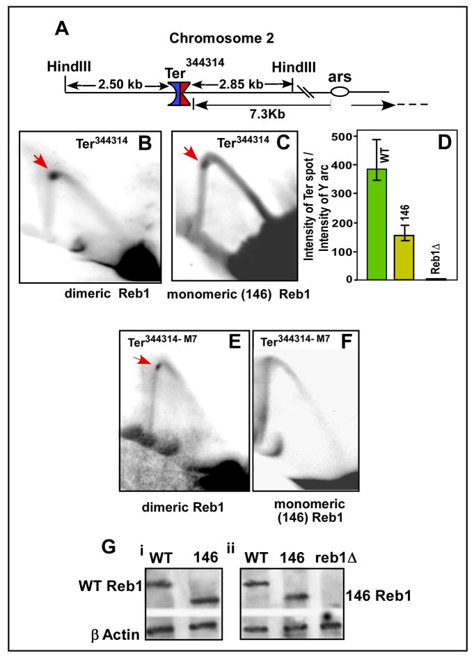
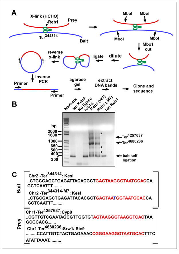
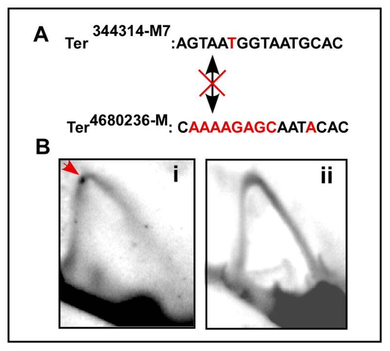
DNA transactions driven by long-range protein-mediated inter- and intrachromosomal interactions have been reported to influence gene expression. Here, we report that site-specific replication termination in Schizosaccharomyces pombe is modulated by protein-mediated interactions between pairs of Ter sites located either on the same or on different chromosomes. The dimeric Reb1 protein catalyzes termination and mediates interaction between Ter sites. The Reb1-dependent interactions between two antiparallel Ter sites in cis caused looping out of the intervening DNA in vitro and enhancement of fork arrest in vivo. A Ter site on chromosome 2 interacted pairwise with two Ter sites located on chromosome 1 by chromosome kissing. Mutational inactivation of the major interacting Ter site on chromosome 1 significantly reduced fork arrest at the Ter site on chromosome 2, thereby revealing a cooperative mechanism of control of replication termination.


Figures

Fig. 1
Fig. 1. Locations of the Ter sites of S. pombe in rDNA and of Reb1 terminator protein in the nucleoli
A, schematic representation of the nontranscribed spacer region of rDNA, the 3 Ter sites and the fork pausing site RFP4 are shown, Ter1 binds to Sap1 and Ter2 and Ter3 bind to Reb1 proteins, Ter2 and Ter3 arrest replication forks approaching from one direction (red arrows), and transcription catalyzed by RNA polymerase I from the opposite direction (blue arrows); B, schematic representation of Reb1 protein showing the N-terminal dimerization domain in green and the predicted myb1 domains in red, the myb associated domain; the region of Reb1 from the amino acid residues 156 to 418 is able to arrest replication forks; C, (i) micrographs showing a field of cells in phase contrast, the same field illuminated at a wavelength that shows red fluorescence for Gar2, a nucleolar protein, yellow-green fluorescence emitted by Reb-GFP and a merged image of Gar2-cherry and Reb1-GFP shown in the panels (ii), (iii) and (iv) respectively.
Fig. 2
Fig. 2. In vitro Reb1-mediated ligation enhancement assay showing that Reb1 promotes interactions between not only two WT Ter sites but also between a WT and a nonfunctional mutant Ter site M7 in vitro
A, sequences showing the WT, canonical Ter and the M7 mutant form with the G to T transversion marked with a red dot; B, a schematic representation of the assay method, the plasmid pSW1 contains either two WT Ter sites or a WT and a M7 site placed on either sides of the unique EcoR1 site, DNA looping caused by the binding of the WT Reb1 but not a truncated, monomeric mutant for is manifested by enhancement of DNA circularization of the linearized, labeled DNA that is measured by counting gene 6 exonuclease resistant label; C, Ligation enhancement kinetics showing that two WT sites loop DNA in the presence of the WT Reb1 but not when the truncated form of the protein was supplied, controls show no enhancement of ligation kinetics when a single Ter site was incubated with either the WT or the truncated form of Reb1 or when only ligase was added to the reaction mixture containing the DNA with two WT sites but no reb1 protein; D, ligation kinetics showing that both the substrate containing either two wt sites or a wt site and a M7 site yield approximately equal amounts of enhancement of ligation rate; E, measurement of ligation kinetics showing that a DNA substrate with one WT site and a M7 site show significant enhancement of ligation rate when incubated with the WT Reb1 but not with the mutant monomeric form of Reb1 protein, control experiments using the monomeric protein and DNA substrates with either two or a single WT Ter site did not show more than background levels of ligation enhancement; F, ligation kinetics showing that placing the WT and M7 sites in a mutually parallel orientation does not affect DNA looping.
Fig. 3
Fig. 3. Autoradiogram of a 6% polyacrylamide nondenaturing gel showing exonuclease III footprints of WT Reb1 supporting Ter-Ter interaction in vitro
A, schematic representation of the exonuclease III footprinting experiments, the plasmid pSW1 contains a WT Ter site and a mutant M7 site placed on either side of the unique EcoR1 site, protection of the M7 site caused by dimeric Reb1 protein-mediated site-site interaction was measure by the protection of the unique Pst1 site located immediately upstream of the M7 site that generated the ~130bp diagnostic labeled DNA fragment; B, the labeled DNA substrates (25 pmole, each) were incubated with 0.5X (125pmoles), 10X (250 pmoles), 20X, 50X, 100X and 200X wt Reb1 protein and the truncated, monomeric Reb1 of the same range of concentrations, the substrate DNA (25 pmoles) digested with exo III to completion and resolved by electrophoresis. The arrow shows the protected fragment diagnostic of WT Ter – M7 interaction; the truncated Reb1 protein did not generate any protected fragment excepting at the two highest concentration of the protein (100X and 200X) that showed trace amounts of the fragment; C, autoradiogram showing that the control substrate containing a single WT Ter site did not generate any protected fragment when bound to either the WT or the truncated form of Reb1. The location of the protected fragment is marked by an arrow in the “marker” lane; D, autoradiogram showing the extent of protection of the solo M7 region in the presence of WT Reb1 of the same concentration range as in 3B. See also Fig. S1.
Fig. 4
Fig. 4. Representative images of 2D gels of replication intermediates of various DNA substrates
A, schematic diagram of the plasmid pIRT2 WT-M7 containing an ARS of S. pombe, Leu2 marker, a nonfunctional M7 Ter site and a WT Ter site located 1.3 kb away from the M7 and 2 PvuII sites; the diagram also shows that the dimeric WT Reb1 is expected to promote looping interaction between the M7 and the WT site located in an antiparallel orientation; B, i–iv, images of 2D gels of DNA substrates containing a single WT site, with a single M7 site, containing both the M7 and the WT site in cells expressing the WT Reb1 and the same M7-WT substrate but extracted from cells expressing the truncated, monomeric form of Reb1, respectively; C, diagram of pIRT2-M7-WT-b that contained the two Ter sites separated by 250bp; D, a representative image of a 2D gel of the plasmid shown in C; E, diagram of the pIRT2-M7-TW plasmid with the two Ter sites present in a parallel orientation; F, image of a 2D gel of replication intermediates of the plasmid shown in E; G, diagram of pIRT2-7M-WT plasmid (inverted M7 site) and H, 2D gel pattern of the plasmid shown in G. With the exception of B, iv, in which the replication intermediates were isolated from cells expressing the monomeric Reb1, all of the other samples were extracted from cells expressing the WT reb1 protein. See also Fig. S2.
Fig. 5
Fig. 5. Two dimensional gels of replication intermediates showing that Ter-Ter interactions occur in vivo at natural Ter sites to enhance fork arrest
A, the location of Ter344314 in chromosome II showing the HinD III sites used to cleave the replication intermediates for 2D gel analysis; B, image of a representative 2D gel, of intermediates purified from cells expressing the WT Reb1, showing fork arrest at the Ter; C, the same as in B excepting that the cells were expressing the monomeric form of Reb1; D, quantification of the replication intermediates with standard deviation bars showing that fork arrest at Ter344314; E, a representative image of a 2D gel of fork arrest at at Ter344314-M7 in cells expressing WT Reb1; F, 2D gel pattern of Ter344314-M7 in cells expressing the truncated monomeric Reb1 protein; note that fork arrest was abolished in cells expressing the monomeric Reb1; G, Western blots showing expression of WT Reb1 and 146 Reb1 in cells containing the normal (Gi) and the M7 form of Ter344314; the blots were developed using polyclonal antibodies raised against Reb1, the loading control is β tubulin. Arrows show termination spots.
Fig. 6
Fig. 6. Identification of sequences interacting with Ter344314 by 4C analysis
A, schematic diagram showing the major steps of 4C analysis, the key enzymes/manipulation used at each step are indicated (see the text for further explanation); B, representative agarose gel of the inverse PCR products from a representative 4C analysis that used Ter344314 and Ter344314-M7 as baits, the asterisks show faint minor products that were found to be nonspecific PCR products, the lanes marked Reb1 (WT) and Reb1 (M7) refer to products from cells expressing WT Reb1 protein and which contained the Ter344314 and Ter344314-M7 bait sequences respectively; C, sequences of the bait and the prey sites; the two prey sequences captured by 4C namely Ter4680236 and Ter4257637 are both located in chromosome I, the third major band was self ligated bait sequence which, as expected, is missing from the ligase minus control lane in B (the primer sequences are shown in supplementary data).
Fig. 7
Fig. 7. 2D gel analysis showing that mutational knockout of Ter4680236 on chromosome 1 abolishes fork arrest at Ter344314-M7 on chromosome 2
A, sequences of the mutated Ter4580236-M and Ter344314-M7, the altered bases are shown in red; B, images of representative 2D gels showing fork arrest (red arrow) at the control M7 site (Bi) and abolition of the same in cells harboring Ter2680436-M (Bii). The crossed double headed arrow indicates that the two sites do not interact with each other because Ter4580236-M no longer binds to Reb1.

Similar articles

Cited by

References

    1. Arcangioli B, Klar AJ. A novel switch-activating site (SAS1) and its cognate binding factor (SAP1) required for efficient mat1 switching in Schizosaccharomyces pombe. Embo J. 1991;10:3025–3032. - PMC - PubMed
    1. Bastia D, Mohanty BK. Mechanisms for completing DNA replication. In: DePamphilis M, editor. DNA Replication in Eukaryotic Cells. Plainview, NY: Cold Spring harbor Laboratory Press; 1996. pp. 177–215.
    1. Bastia D, Mohanty BK. Termination of DNA Replication. In: DePamphilis M, editor. DNA replication and human disease. Cold Spring Harbor, New York: Cold Spring Harbor Laboratory Press; 2006. pp. 155–174.
    1. Bastia D, Zzaman S, Krings G, Saxena M, Peng X, Greenberg MM. Mechanism of replication termination as revealed by Tus-mediated polar arrest of a sliding helicase. Proc Natl Acad Sci USA. 2008;105:12831–12836. - PMC - PubMed
    1. Biswas S, Bastia D. Mechanistic insights into replication termination as revealed by investigations of the Reb1-Ter3 complex of Schizosaccharomyces pombe. Molecular & Cellular Biology. 2008;28:6844–6857. - PMC - PubMed

Publication types

MeSH terms