. 2010 Sep 17;142(6):889-901.
doi: 10.1016/j.cell.2010.08.017.

Membrane remodeling induced by the dynamin-related protein Drp1 stimulates Bax oligomerization

Affiliations

Membrane remodeling induced by the dynamin-related protein Drp1 stimulates Bax oligomerization

Sylvie Montessuit et al. Cell. .

Abstract

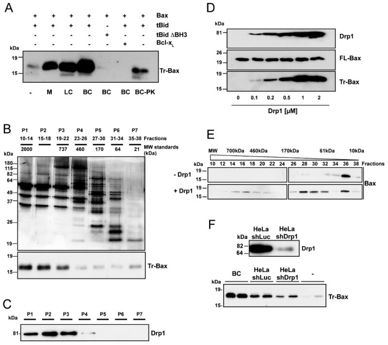
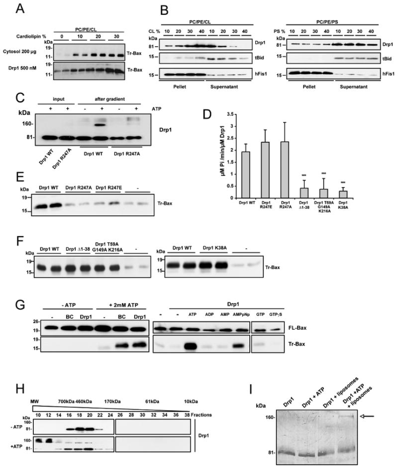
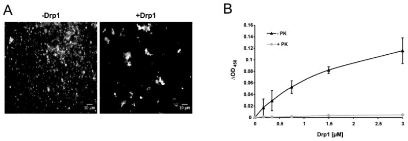
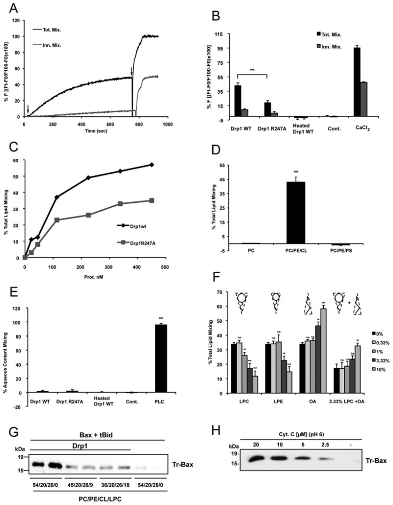
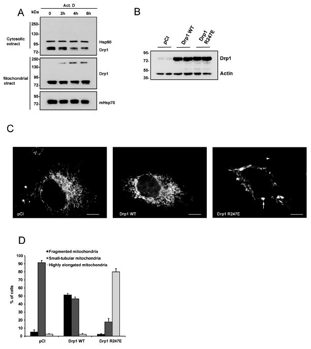
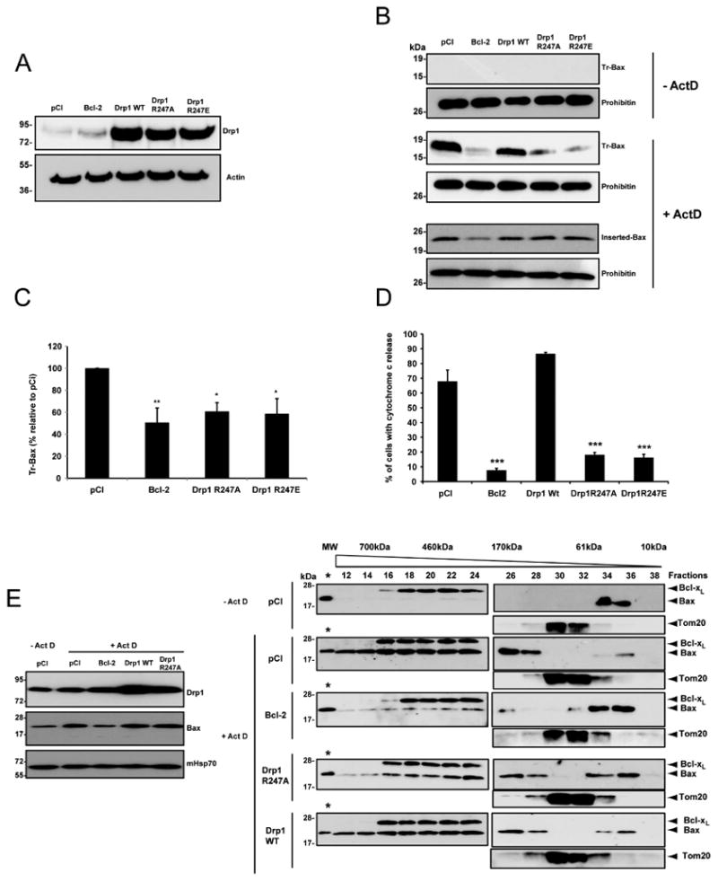
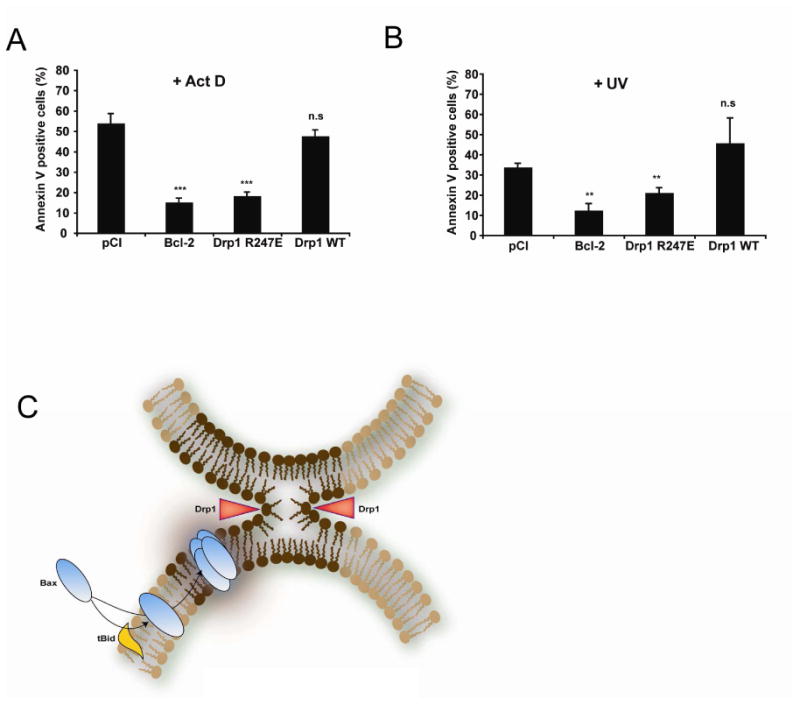
In response to many apoptotic stimuli, oligomerization of Bax is essential for mitochondrial outer membrane permeabilization and the ensuing release of cytochrome c. These events are accompanied by mitochondrial fission that appears to require Drp1, a large GTPase of the dynamin superfamily. Loss of Drp1 leads to decreased cytochrome c release by a mechanism that is poorly understood. Here we show that Drp1 stimulates tBid-induced Bax oligomerization and cytochrome c release by promoting tethering and hemifusion of membranes in vitro. This function of Drp1 is independent of its GTPase activity and relies on arginine 247 and the presence of cardiolipin in membranes. In cells, overexpression of Drp1 R247A/E delays Bax oligomerization and cell death. Our findings uncover a function of Drp1 and provide insight into the mechanism of Bax oligomerization.


Figures

Figure 1
Figure 1. Drp1 promotes Bax oligomerization in the presence of tBid
(A) PC/PE/CL (54/20/26 mol%) liposomes were incubated with the indicated proteins at 30°C for 30 min before ultracentrifugation, resuspension in KCl buffer and incubation with trypsin for 2 h at 30°C. Trypsin-resistant Bax (Tr-Bax) was analyzed by immunoblotting. M: salt-extracted liver mitochondrial proteins (400 μg); LC: rat liver cytosolic extract (200 μg); BC: rat brain cytosolic extract (200 μg); BC-PK: BC (200 μg) treated with proteinase K. Bax (50 nM); tBid (10 nM); tBid ΔBH3 (10 nM); Bcl-xL (1 μM). The blot is representative of three independent experiments. (B) Purification steps of Bax-activating proteins: silver stained SDS-polyacrylamide gel electrophoresis of BC proteins fractionated by size exclusion chromatography (top panel); Bax-activating proteins (assessed as in A) are found in P1-P3 fractions (lower panel). Blots are representative of at least three independent experiments. P1 displayed the highest specific BAF activity and was found to contain GAPDH, Aldolase, Tubulin, Microtubule Associated Protein 1, Dynein, Spectrin, Actin, Gelsolin, Alpha Actinin, Cofilin and Drp1. Except Drp1, all proteins were found to be inactive in the Bax oligomerization assay. (C) Immunoblot showing that Drp1 is present in P1-P3 fractions. (D) Dose-response analysis of in vitro tBid-induced Bax oligomerization with increasing concentrations of recombinant Drp1. PC/PE/CL liposomes were incubated with 10 nM tBid and 50 nM Bax and increasing amounts of recombinant Drp1 before trypsin digestion and Bax analysis. Upper immunoblots show levels of Drp1 and Bax before trypsin digestion. The blot is representative of three independent experiments. (E) Analysis of tBid-induced Bax oligomerization in isolated liposomes in the absence or presence of 1 μM Drp1 by size exclusion chromatography. PC/PE/CL liposomes were incubated with 10 nM tBid, 50 nM Bax and 1 μM Drp1. Liposomes were then lysed in 2% CHAPS, 200 nM NaCl, and proteins fractionated by size exclusion chromatography. The elution profile of Bax was analyzed by immunoblotting. (F) Upper panel: Western blot analysis of Drp1 in control (shLuc) and Drp1-depleted (shDrp1) HeLa cells. Lower panel: analysis of Bax activating capacity of control and Drp1-depleted HeLa cytosolic extracts (200 μg each). Brain cytosol (BC) was also tested at 200 μg. In each experiment, the same cytosolic extract from HeLa cells was tested in duplicate. The experiment was repeated twice, using the same cell lines but with a different cytosolic extract preparation.
Figure 2
Figure 2. Drp1 acts independently of its GTPase activity, requires ATP, and its binding to cardiolipin is essential for tBid-induced Bax oligomerization
(A) PC/PE/CL liposomes were prepared with increasing concentrations of CL and concomitant reduction of phosphatidylcholine. They were incubated with 10 nM tBid and 50 nM Bax and with 200 μg brain cytosol or 500 nM Drp1 before trypsin digestion and Bax analysis. This blot is representative of three independent experiments. (B) Preferential binding of Drp1 and tBid to CL. Liposomes with increasing amounts of CL (PC/PE/CL, left panel) or PS (PC/PE/PS, right panel) were incubated in the presence of Drp1 (1 μM) or tBid (10 nM), centrifuged and analyzed by Western blotting for the presence of Drp1 or tBid in the pellet or the supernatant. As a control, liposomes were incubated with the integral membrane protein hFis1. Each panel represents a separate experiment. This blot is representative of two independent experiments. See also Figure S1. (C) PC/PE/CL (54/20/26) liposomes were incubated with Drp1 R247A or Drp1 WT (500 nM each) alone in the presence or absence of ATP and loaded on a sucrose gradient before ultracentrifugation. Drp1 R247A and Drp1 WT were analyzed by immunoblotting in the suspension before gradient centrifugation and after centrifugation in the floating liposomal suspension. This blot is representative of three independent experiments. (D) GTPase activity of Drp1 WT and Drp1 mutants (1 μM each). Values are means of three independent experiments ± SD. Drp1 GTPase mutants differed from WT with ***: p< 0.001. (E) Comparison of the effects of Drp1 WT, Drp1 R247A and Drp1 R247E (1 μM each) on tBid-induced Bax oligomerization. PC/PE/CL liposomes were incubated in the presence of 10 nM tBid, 50 nM Bax and the indicated proteins before trypsin digestion and Bax analysis. The blot is representative of three independent experiments. See also Figure S2. (F) Analysis of Drp1 GTPase mutants (1 μM each) for their capacity to stimulate tBid-induced Bax oligomerization. Blots show duplicates and are representative of three independent experiments. (G) In vitro analysis of tBid-induced Bax oligomerization in the absence or presence of ATP (left blot) or in the presence of ATP or other nucleotides, each tested at 2.5 mM (right blot). Experiments were performed in the presence or absence of 1 μM Drp1 or 200 μg brain cytosol (BC) (right blot; the two parts of this blot correspond to the same membrane and identical exposure times). Upper blots show Bax binding to liposomes. Blots are representative of at least three independent experiments. (H) Analysis of Drp1 by size exclusion chromatography. Liposomes were incubated with Drp1 (1 μM), Bax (50 nM) and tBid (10 nM) in the presence or absence of 2.5 mM ATP, solubilized with CHAPS, separated by size exclusion chromatography and the elution profile of Drp1 was analyzed by immunoblotting. (I) Drp1 (1 μM) was incubated with liposomes and/or 2.5 mM ATP, run on a polyacrylamide gel under reducing conditions and detected by Coomassie staining after transfer to a nitrocellulose membrane. The arrow points to Drp1 dimers.
Figure 3
Figure 3. Drp1 triggers membrane tethering
(A) Fluorescence microscopy images of PC/PE/CL liposomes containing 1% NBD-PE in the presence of ATP and the presence or absence of 1 μM Drp1. (B) Dose-response of liposome aggregation in the presence of increasing concentrations of Drp1 (black line). Proteinase K was added to aggregated liposomes and the turbidity of the liposome suspension was measured 5 min later (grey line). Turbidity of the suspension was measured at 450 nm. ΔOD values represent the difference between OD450 of the liposome suspension incubated in the presence of proteins and OD450 values in the absence of proteins. Results are means of three independent experiments ± SD.
Figure 4
Figure 4. Drp1 potentiates tBid-induced Bax oligomerization by promoting hemifusion of cardiolipin-containing membranes
(A) Representative time courses of total lipid mixing (Tot. Mix.) and inner monolayer lipid mixing (Inn. Mix.) induced by 225 nM Drp1 WT in PC/PE/CL LUVs. Lipid mixing was monitored by the NBD/Rhodamine lipid dilution assay. For inner monolayer lipid mixing NBD + Rho-liposomes were treated with appropriate amounts of sodium dithionite to quench NBD fluorescence of the outer leaflet. Unlabeled and NBD + Rho-Labeled liposomes were mixed and incubated for 5 min before protein addition. The extent of lipid mixing was quantified on a percentage basis according to the equation: (Ft − F0/F100 − F0) × 100 where Ft is the measured fluorescence of protein-treated LUVs at time t, F0 is the initial fluorescence of the LUV suspension before protein addition, and F100 is the fluorescence value after complete disruption of LUVs by addition of 10 mM β-octylglucoside (OG). First arrow shows time of addition of Drp1 WT to the liposome suspension and the second arrow denotes addition of OG. (B) Comparison of the degree of total lipid mixing and inner monolayer lipid mixing elicited by Drp1 WT, Drp1 R247A and heat-denatured Drp1 WT (Heated Drp1 WT) and 10 mM CaCl2. As a control, Drp1 WT was incubated with NBD/Rho containing LUVs but in the absence of unlabeled LUVs (Cont.). 100% total lipid mixing value corresponds to LUVs treated with OG. Mean values ± S.E. are shown for three to seven independent experiments. Drp1 WT differed from Drp1 R247A with: *** p < 0.001. See also Figure S3. (C) Dose response of Drp1 WT and Drp1 R247A on total lipid mixing. (D) Lipid mixing elicited by Drp1WT in LUVs containing either PC (100), PC/PE/CL (54/20/26) or PC/PE/PS (28/20/52). Mean values ± S.E. are shown for three independent experiments. PC and PC/PE/PS differed from PC/PE/CL with *** p < 0.001. (E) Comparison of the degree of aqueous content mixing elicited by Drp1 WT, Drp1 R247A, heat-denatured Drp1 WT (Heated Drp1 WT) and 0.1 nM Phospholipase C from Bacillus cereus (PLC). Content mixing was monitored by ANTS/DPX aqueous content mixing assay; 100% of content mixing was determined using LUVs containing both 12.5 mM ANTS and 45 mM DPX. Drp1 WT was incubated with ANTS containing LUVs as a control (Cont.). Mean values ± S.E. are shown for three independent experiments. Drp1 WT, Drp1 R247A, and heated Drp1 WT differed from PLC with: *** p < 0.001. (F) Total lipid mixing induced by 158 nM Drp1WT in PC/PE/CL vesicles upon external addition of indicated concentrations of LPC, LPE and OA. Mean values ± S.E. are shown for three to seven independent experiments. Each LPC, LPE, OA concentration differed from 0% with **: p < 0.01. ***: p < 0.001 or was not significantly different (ns). (G) PC/PE/CL liposomes containing 9 or 18 mol% LPC were used in in vitro Bax oligomerization assays. Drp1 was used at 1 μM. Blot shows duplicates and is representative of three independent experiments. (H) PC/PE/CL liposomes were incubated with tBid and Bax and increasing concentrations of cytochrome c at pH 6 before assaying Bax oligomerization. The blot is representative of three independent experiments.
Figure 5
Figure 5. Drp1 oligomerization during apoptosis and impact of Drp1 247A/E mutants on mitochondrial morphology
(A) Mitochondrial and cytosolic extracts were prepared from HeLa cells cultured in the absence (time 0) or in the presence of ActD (3 μM) for 2, 4 and 6 h. Cell extracts were separated by SDS-PAGE in the absence of DTT and analyzed for Drp1 by Western blotting. Hsp90 and mHsp70 were used as loading controls for cytosolic and mitochondrial extracts respectively. The blots are representative of four independent experiments. (B-D): HeLa cells were transfected with an empty DNA vector (pCI) or with plasmids encoding Drp1 WT or Drp1 R247E together with a mitochondria-targeted YFP. 48h later Drp1 expression levels were quantified in total cell extracts by Western blot using an antibody to Drp1. Actin was used as a loading control. In parallel, mitochondrial morphology was analyzed by fluorescence microscopy. (B) Immunoblot shows that cells transfected with plasmids encoding Drp1 WT or Drp1 R247E expressed equivalent amounts of Drp1 WT and Drp1 R247E proteins. Actin was used as a loading control. See also Figure S4. (C) Morphology of mitochondria in HeLa cells observed by fluorescence microscopy. White arrow denotes the presence of vesicular dilatations while arrowhead denotes highly elongated mitochondria. Bar is 5 μm (D) Quantification of mitochondrial morphology. Mitochondria were divided into three classes: small tubular, which corresponds to cells mainly filled with small filaments of ∼2 μm or less; fragmented that corresponds to punctiform mitochondria; highly elongated, which corresponds to cells mainly filled with mitochondria > 5μm.
Figure 6
Figure 6. Expression of Drp1 R247A/E mutants decreases cytochrome c release and Bax oligomerization in response to apoptotic stimuli
(A) In all experiments performed to assess cytochrome c release, Bax oligomerization, and cell death (Figure 7), HeLa cells were transfected with an empty vector (pCI) or with plasmids encoding Bcl-2, Drp1 WT or Drp1 R247A/E. The efficacy of transfection varied between 60 to 80%. Drp1 WT and Drp1 R247A/E were overexpressed at equivalent levels as shown by the immunoblot perfomed with an antibody to Drp1. Actin was used as a loading control. (B, C) Analysis of Bax insertion in the outer mitochondrial membrane by alkali treatment and Bax oligomerization by the trypsin digestion assay. HeLa cells were transfected as indicated in (A). 72 h after transfection, apoptosis was induced with 3 μM ActD (+ ActD). 4 h later, mitochondria were isolated and analyzed for Bax insertion by alkali treatment (inserted Bax) or Bax oligomerization by trypsin digestion (Tr-Bax). Tr-Bax was also analyzed in untreated HeLa cells (-ActD). Prohibitin was used as a mitochondrial loading control. Note that although Bax insertion was similar in cells transfected with pCI, Drp1 WT or Drp1 R247A/E vectors, Tr-Bax levels were significantly lower in cells expressing Drp1 R247A/E or Bcl-2 as shown in (C) which is a compilation of results from four different experiments performed for pCI, Bcl-2, Drp1 R247A and Drp1 R247E. Results represent a quantification of the immunoblots for trypsin-resistant Bax. Data are mean ± SE (n = 6); *p < 0.05, ** p < 0.01. (D) HeLa cells were transfected as indicated in (A). 72 h later cells were treated with ActD for 4 h in the presence of 100 μM z-VAD to avoid their detachment from the culture dish, and immunostained for cytochrome c. Cells were analyzed by fluorescence microscopy and the number of cells with diffused cytochrome c staining was counted. Histograms display the means of three independent experiments ± S.E; ***: p < 0.001 (E) Analysis of Bax oligomerization by size exclusion chromatography. HeLa cells were transfected as indicated in (A). 72 h after transfection, cells were treated with 3 μM ActD to induce apoptosis. 6h later mitochondria were isolated from cells undergoing apoptosis and from pCI transfected cells that were not exposed to ActD, as a control. Proteins were extracted from membranes with 2% CHAPS in 200 mM NaCl and fractionated by size exclusion chromatography. Proteins in the eluted fractions were precipitated with tricholoroacetic acid and separated by SDS-PAGE before Bax immunoblotting (Right panel). 1 ng recombinant Bax was loaded on every blot (labeled with an asterisk) to control that every blot was exposed similarly to X ray film during the chemiluminescence detection of proteins, allowing direct comparison of all immunoblots. In addition, Bcl-xL and Tom 20 were used as loading controls. The left panel shows levels of Bax that were associated with mitochondria before extraction. Results confirm those shown in (B), lower panel). Drp1 and mHsp70 (used as a loading control) were also blotted. See also Figure S5.
Figure 7
Figure 7. Expression of Drp1 R247A/E delays cell death
(A, B) HeLa cells were transfected with an empty vector (pCI) or with plasmids encoding Bcl-2, Drp1 WT or Drp1 R247E. 72 h later, cell cultures were treated with 3 μM Act D (A) or UV- irradiated (60 mJ/cm2) (B) and apoptosis quantified 6 h later by Annexin V staining and FACS analysis. Values are the average of six independent experiments ± S.E. **: p < 0.01; ***: p < 0.001; (n.s.), not significant. (C) Model explaining the role of mitochondrial membrane hemifission or hemifusion intermediates in Bax oligomerization. During apoptosis, Bax is recruited to the outer mitochondrial membrane by tBid (or other BH3 only proteins) where it inserts. At the same time, Drp1 constricts the organelle as indicated by red arrowheads, triggering the formation of a hemifission intermediate. This membrane remodeling (dark brown part of the membrane) promotes Bax oligomerization. We speculate that contact sites between the inner and outer membranes, that are enriched in CL, could also be privileged sites for the formation of hemifusion intermediates, independently of Drp1. Contact sites could therefore represent additional sites in which Bax oligomerization would occur.

Similar articles

Cited by

References

    1. Ardail D, Privat JP, Egret-Charlier M, Levrat C, Lerme F, Louisot P. Mitochondrial contact sites. Lipid composition and dynamics. J Biol Chem. 1990;265:18797–18802. - PubMed
    1. Arnoult D, Rismanchi N, Grodet A, Roberts RG, Seeburg DP, Estaquier J, Sheng M, Blackstone C. Bax/Bak-dependent release of DDP/TIMM8a promotes Drp1-mediated mitochondrial fission and mitoptosis during programmed cell death. Curr Biol. 2005;15:2112–2118. - PubMed
    1. Basanez G, Fidelio GD, Goni FM, Maggio B, Alonso A. Dual inhibitory effect of gangliosides on phospholipase C-promoted fusion of lipidic vesicles. Biochemistry. 1996;35:7506–7513. - PubMed
    1. Basanez G, Sharpe JC, Galanis J, Brandt TB, Hardwick JM, Zimmerberg J. Bax-type apoptotic proteins porate pure lipid bilayers through a mechanism sensitive to intrinsic monolayer curvature. J Biol Chem. 2002;277:49360–49365. - PubMed
    1. Bellot G, Cartron PF, Er E, Oliver L, Juin P, Armstrong LC, Bornstein P, Mihara K, Manon S, Vallette FM. TOM22, a core component of the mitochondria outer membrane protein translocation pore, is a mitochondrial receptor for the proapoptotic protein Bax. Cell Death Differ. 2006;14:785–794. - PubMed

Publication types