. 2010 Sep 17;142(6):902-13.
doi: 10.1016/j.cell.2010.08.029.

Influenza virus M2 protein mediates ESCRT-independent membrane scission

Affiliations

Influenza virus M2 protein mediates ESCRT-independent membrane scission

Jeremy S Rossman et al. Cell. .

Abstract

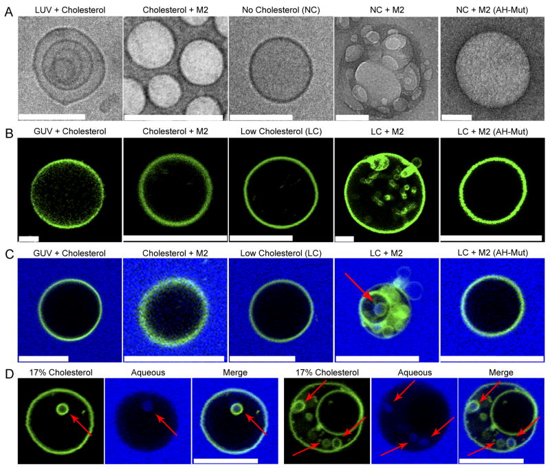
Many viruses utilize host ESCRT proteins for budding; however, influenza virus budding is thought to be ESCRT-independent. In this study we have found a role for the influenza virus M2 proton-selective ion channel protein in mediating virus budding. We observed that a highly conserved amphipathic helix located within the M2 cytoplasmic tail mediates a cholesterol-dependent alteration in membrane curvature. The 17 amino acid amphipathic helix is sufficient for budding into giant unilamellar vesicles, and mutation of this sequence inhibited budding of transfected M2 protein in vivo. We show that M2 localizes to the neck of budding virions and that mutation of the M2 amphipathic helix results in failure of the virus to undergo membrane scission and virion release. These data suggest that M2 mediates the final steps of budding for influenza viruses, bypassing the need for host ESCRT proteins.


Figures

Figure 1
Figure 1. Conservation of the M2 Amphipathic Helix
(A) Diagram of structural motifs in the M2 protein. The TM domain/ion channel pore, CRAC motif and amphipathic helix are indicated. Alanine substituted residues in the M2(AH-Mut) protein are shown in red. (B) Helical wheel plot of the M2 amphipathic helix is shown as generated at http://heliquest.ipmc.cnrs.fr. Hydrophilic residues are shown in black and hydrophobic resides in grey. The red line separates the two faces of the helix. See also Fig. S1.
Figure 2
Figure 2. M2 Alters Membrane Curvature in a Cholesterol-Dependent Manner
(A) LUVs or cholesterol-free LUVs were reconstituted in the presence or absence of 100 μg purified M2 or M2(AH-Mut) protein and analyzed by cryo-electron microscopy. Scale bars indicate 100 nm. (B) LUVs prepared as above, with the addition of a fluorescent membrane dye (shown in green), were dehydrated, electroformed into GUVs and immediately imaged. (C) GUVs, prepared as above, with 50 μg purified M2 or 100 μg M2(AH-Mut) protein were resuspended with 0.5 mg/ml of lucifer yellow (shown in blue). NC indicates LUVs containing no cholesterol. LC indicates GUVs containing 0.5 molar % cholesterol. (D) Two examples of M2-containing GUVs, prepared as above, containing 17 molar % cholesterol. Arrows indicate lucifer yellow containing ILVs. Scale bars indicate 10 μm. See also Figs. S2, S3 and Movies S1, S2.
Figure 3
Figure 3. The M2 Amphipathic Helix Causes Membrane Budding in vitro
(A) GUVs electroformed with 30 or 0.5 molar % of cholesterol were treated with 10 μM of the indicated peptide and imaged within 1 h. (B) GUVs were prepared and treated as above except that 0.5 mg/ml of lucifer yellow (shown in blue) was added to the resuspension buffer. (C) GUVs, prepared as above, were treated with M2AH peptide at the indicated concentrations for 1 h and imaged. (D) GUVs, prepared as above with the indicated molar % of cholesterol, were treated with 10 μM of M2AH peptide for 1 h and imaged. 17% cholesterol LY-post scission indicates 17% cholesterol GUVs to which lucifer yellow was added 1 h post-treatment with 10 μM of M2AH peptide. (E) GUVs, prepared as above (with the exception of M2AH-TMR treated samples, for which lucifer yellow was omitted from the resuspension buffer), were treated with 10 μM of the indicated peptide and imaged within 1 h. The M2AH-WSN peptide corresponds to the M2 amphipathic helix of A/WSN/33, M2AH-SOIV to A/California/05/2009, M2AH-Sc to a scrambled sequence of the M2AH peptide, M2AH-TMR to rhodamine-labled M2AH peptide. LC indicates GUVs containing 0.5 molar % of cholesterol. Scale bars indicate 10 μm. See also Figs. S3, S4 and Movie S3.
Figure 4
Figure 4. The M2 Amphipathic Helix Causes Membrane Budding at Phase Boundaries
Phase-separated GUVs, electroformed with 20 or 5 molar % of cholesterol and fluorescent markers for the Lo and Ld phase, were treated as indicated and imaged within 1 h. (A) Representative example of phase-separated GUV containing 20 molar % of cholesterol. (B) Phase-separated GUVs were prepared as above with the Lo marker omitted. GUVs were treated with 10 μM of the M2AH-TMR peptide for 1 h and imaged. Arrows indicate clustered M2AH peptide at the phase boundary. (C) Phase-separated GUVs prepared as in Fig. 4a containing 20 molar % cholesterol or (D) 5 molar % were resuspended with lucifer yellow, treated with 10 μM of the M2AH peptide for 1 h and imaged. Ld phase is shown in red, the Lo phase in green and the aqueous media in blue. (E) Time lapse images of a GUV from Fig. 4d. Note: loss of the Ld signal in later time points is due to the rapid bleaching of the red signal. (F) Plasma membrane spheres were prepared from wt M2 expressing cells or (G) M2(AH-Mut) expressing cells, GM1 was crosslinked to mark the Lo phase (green) and spheres were stained for M2 (red). Scale bars indicate 10 μm. Arrows indicate sites of budding. See also Fig. S5 and Movie S4.
Figure 5
Figure 5. M2 Causes Membrane Budding in vivo
(A) 293T cells were transfected with M2 or M2(AH-Mut) expression plasmids and 24 h post-transfection the supernatant was harvested, concentrated, and M2 content was analyzed by western blot as compared to whole cell lysates. Values indicate average +/− standard deviation of the percentage of M2 found in the supernatant and calculated from a minimum of three repeats. M2-low indicates cells transfected with a 4-fold reduction of DNA to provide comparable M2 expression levels to that of M2(AH-Mut). The vertical line indicates separate blots. (B) Concentrated supernatant from Fig. 5a was analyzed by electron microscopy. Scale bars indicate 100 nm. The inset is an enlargement of a 100nm square region of the panel below.
Figure 6
Figure 6. M2 Localizes to the Neck of Budding Virions
MDCK cells were infected with an MOI of 3 pfu/cell of A/Udorn/72 for 18 h. (A) Virus-infected cells were fixed and processed for immunofluorescent detection of HA (shown in green) and M2 (shown in red). (B) Magnification of images shown in Fig. 6a. Scale bars indicate 10 μm. (C) Virus-infected cells were stained for M2 via immuno-gold labeling and thin sections were analyzed by electron microscopy. (D) Magnification of images shown in Fig. 6c. Scale bars indicate 100 nm. Arrows indicate M2 foci at sites of virus budding. See also Fig. S6.
Figure 7
Figure 7. The M2 Amphipathic Helix is Necessary for Membrane Scission and Virion Release
MDCK cells were infected with an MOI of 3 pfu/cell of (A) A/Udorn/72, (A-C) A/Udorn/72 M2(AH-Mut) or (D) A/Udorn/72-ΔM2 for 18 h and thin sections were analyzed by electron microscopy. M2 was detected via immunogold labeling in (B). Arrows indicate foci of M2 at points of failed membrane scission. Scale bars indicate 100 nm.

Comment in

  • Influenza exits the cell without an ESCRT.
    Göttlinger HG. Göttlinger HG. Cell. 2010 Sep 17;142(6):839-41. doi: 10.1016/j.cell.2010.08.036. Cell. 2010. PMID: 20850005
  • Severing the bud.
    David R. David R. Nat Rev Microbiol. 2010 Nov;8(11):757. doi: 10.1038/nrmicro2458. Nat Rev Microbiol. 2010. PMID: 21080549 No abstract available.

References

    1. Allain JM, Ben Amar M. Budding and fission of a multiphase vesicle. Eur Phys J E. 2006;20:409–420. - PubMed
    1. Antonny B. Membrane deformation by protein coats. Curr Opin Cell Biol. 2006;18:386–394. - PubMed
    1. Barman S, Adhikary L, Chakrabarti AK, Bernas C, Kawaoka Y, Nayak DP. Role of transmembrane domain and cytoplasmic tail amino acid sequences of influenza a virus neuraminidase in raft association and virus budding. J Virol. 2004;78:5258–5269. - PMC - PubMed
    1. Brown DA, Rose JK. Sorting of GPI-anchored proteins to glycolipid-enriched membrane subdomains during transport to the apical cell surface. Cell. 1992;68:533–544. - PubMed
    1. Bruce EA, Digard P, Stuart AD. The rab11 pathway is required for influenza a virus budding and filament formation. J Virol. 2010;84:5848–5859. - PMC - PubMed

Publication types

Substances