. 2010 Oct 1;143(1):71-83.
doi: 10.1016/j.cell.2010.08.038. Epub 2010 Sep 16.

Identification of aneuploidy-tolerating mutations

Affiliations

Identification of aneuploidy-tolerating mutations

Eduardo M Torres et al. Cell. .

Abstract

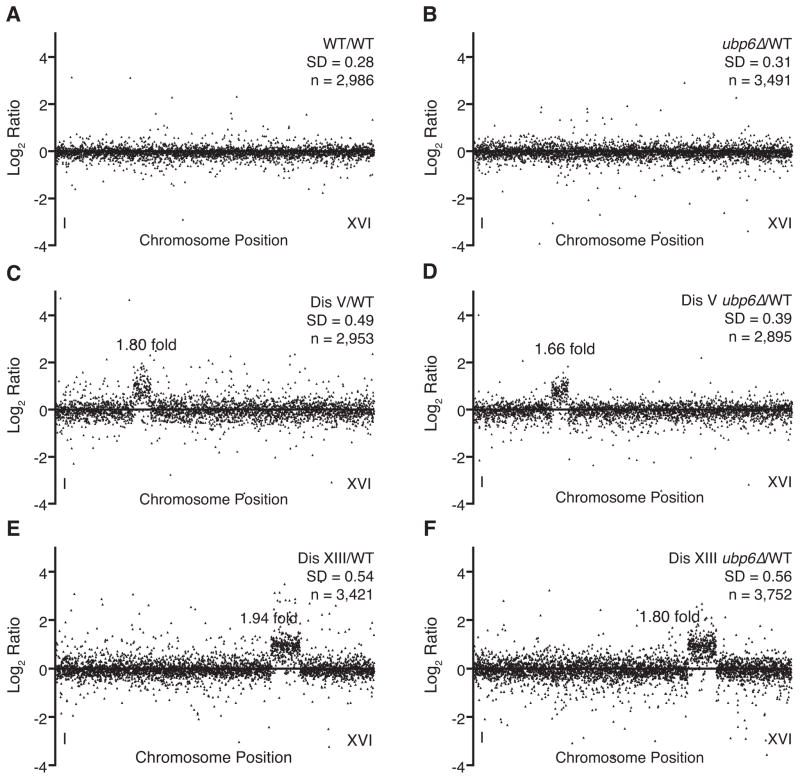
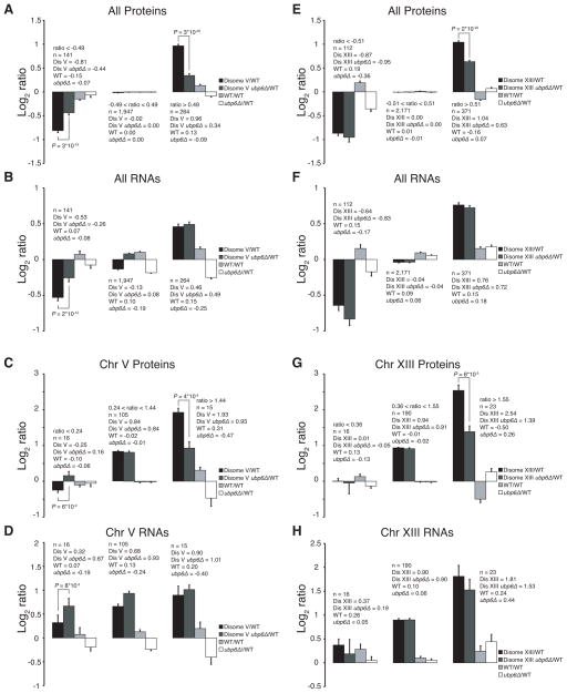
Aneuploidy causes a proliferative disadvantage in all normal cells analyzed to date, yet this condition is associated with a disease characterized by unabated proliferative potential, cancer. The mechanisms that allow cancer cells to tolerate the adverse effects of aneuploidy are not known. To probe this question, we identified aneuploid yeast strains with improved proliferative abilities. Their molecular characterization revealed strain-specific genetic alterations as well as mutations shared between different aneuploid strains. Among the latter, a loss-of-function mutation in the gene encoding the deubiquitinating enzyme Ubp6 improves growth rates in four different aneuploid yeast strains by attenuating the changes in intracellular protein composition caused by aneuploidy. Our results demonstrate the existence of aneuploidy-tolerating mutations that improve the fitness of multiple different aneuploidies and highlight the importance of ubiquitin-proteasomal degradation in suppressing the adverse effects of aneuploidy.


Figures

Figure 1
Figure 1. Evolution of Aneuploid Yeast Strains
(A) Doubling times of disome V (open squares), disome VIII (open triangles), disome XI (open circles), and wild-type cultures (open diamonds) were measured at the indicated times. The arrows indicate the generation when growth rates increased. See also Table S1. (B) Doubling times of wild-type cells (black bar), parental disomes (red bars) and evolved isolates (open bars) were determined in −His+G418 medium at room temperature (n = 3, error bars represent +/− standard deviations (SD), *p value < 0.01 student’s t test). Nomenclature: The Roman numerals describe the identity of the disomic chromosome. The number following the dash indicates when the clone was isolated (after 9 or 14 days of continuous growth), and the number following the period describes the identity of the clone. See also Table S2 and Figure S1. (C) Gene expression analysis of wild-type, parental and evolved disomic strains grown in batch culture, ordered by chromosome position. Experiments (columns) are ordered by the number of the chromosome that is present in two copies. Data were normalized to account for the extra chromosome present in disomic strains. Upregulated genes are shown in red, downregulated ones in green. See also Table S3 and Figure S2.
Figure 2
Figure 2. Loss of UBP6 Function Increases the Fitness of Strains Disomic for Chromosomes V, VIII, IX or XI
(A) Schematic of the Ubp6 domain structure. The N-terminus contains an ubiquitin-like domain (UBL, amino acids 1–83) and the C-terminus harbors the ubiquitin hydrolase domain (amino acids 83–499). The positions of the catalytic cysteine 118 and the two early stop codons at positions 256 and 404 identified in evolved disome V-14.1 and disome IX-14.1, respectively, are shown. (B) The percentage of cells in co-cultures of strains carrying PGK1 fused to GFP (open squares) and strains harboring a C-terminal truncated version of ubp6 (E256X, closed triangles) was determined at the indicated times. All strains were grown in −His+G418 medium. See also Figure S3. (C) The percentage of cells in co-cultures of strains carrying PGK1 fused to GFP (open squares) and strains harboring a UBP6 deletion (ubp6Δ, closed triangles) was determined at the indicated times. All strains were grown in −His+G418 medium. See also Figure S4. (D) Doubling times of the WT, disome V, evolved disome V-14.1, and disome V ubp6Δ strains grown in −His+G418 medium (n = 3, error bars represent +/− SD). See also Figure S5. (E) Doubling times of the WT, disome V, disome VIII, disome IX and disome XI strains either wild-type for UBP6 or carrying a UBP6 deletion grown in YEPD medium (n = 3, error bars represent +/− SD; *p value < 0.01 student’s t test).
Figure 3
Figure 3. Ubiquitin Depletion Is Not Responsible for the Aneuploidy Tolerance Caused by Loss of UBP6 Function
(A) Wild-type, ubp6Δ, disome V and disome V ubp6Δ cells were grown in −His+G418 medium to an OD600 of 1.0 when 100 μg/ml cycloheximide (time = 0 min) was added. Free ubiquitin and ubiquitin conjugates were analyzed by immunoblotting using an anti-ubiquitin antibody at the indicated times. (B) Ubiquitin levels in the presence (+) or absence (-) of 100 μg/ml CuSO4. (C - H) The percentage of cells in co-cultures of strains carrying PGK1 fused to GFP (open squares) and strains harboring a UBP6 deletion (closed triangles) was determined at the indicated times. All strains carry a CUP1-UBI4 multicopy plasmid whose expression was induced with 100 μg/ml CuSO4. The following strains were compared: (C), wild-type and UBP6 deletion cells; (D), disome V PGK1-GFP and disome V ubp6Δ cells; (E), disome XI PGK1-GFP and disome XI ubp6Δ; (F), wild-type and ubp6E256X truncation strains; (G), disome VIII PGK1-GFP and disome VIII ubp6E256X cells; and (H), disome XI PGK1-GFP and disome XI ubp6E256X cells. All strains were grown in −His+G418 medium. See also Figure S6.
Figure 4
Figure 4. Disomic Strains Exhibit an Increased Reliance on the Proteasome for Survival
(A - D) The percentage of cells in co-cultures of strains carrying PGK1 fused to GFP (open squares) and strains harboring a catalytic dead version of UBP6 (ubp6CA, closed triangles) was determined at the indicated times. The following strains were compared: (A), wild-type and ubp6CA cells; (B), disome VIII PGK1-GFP and disome VIII upb6CA cells; (C), disome IX PGK1-GFP and disome IX ubp6CA cells; (D), disome XI PGK1-GFP and disome XI ubp6CA cells. All strains were grown in −His+G418 medium. See also Figure S7. (E) Proliferation capabilities of WT, rpn6-ts, parental disomes and disomes harboring the rpn6-ts allele cells on YEPD medium at 25°C, 30°C and 35°C. 10-fold serial dilutions are shown.
Figure 5
Figure 5. Quantification of the Proteome of Disome V and Disome XIII Strains
The plots show the log2 ratio of the relative protein abundance compared to wild-type. Protein levels are shown in the order of the chromosomal location of their encoding genes. (A): wild-type/wild-type ratios; (B): Δubp6/wild-type ratios; (C), disome V/wild-type ratios; (D), disome V Δubp6/wild-type ratios; (E), disome XIII/wild-type ratios; (F), disome XIII Δubp6/wild-type ratios. SD = standard deviation, n = number of proteins quantified. See also Supplemental Materials. The number in the graphs shows the fold-increase in protein levels of proteins encoded by genes located on the disomic chromosome relative to the rest of the proteome.
Figure 6
Figure 6. Loss of UBP6 Function Preferentially Affects Proteins Overproduced in Disome V and Disome XIII Cells Relative to Wild-type
(A) Comparison of the means of the log2 ratios of relative abundance of proteins. Proteins are binned based on their relative levels in disome V cells. Bin 1 (left bars) contains proteins whose levels are lower than one SD of the mean (ratio < −0.49, n = 141). Bin 2 (middle bars) contains proteins whose levels fall within one SD of the mean (−0.49 < ratio < 0.49, n = 1947) and Bin 3 (right bars) contains proteins whose levels are greater than one SD (ratio > 0.49, n = 264). Only proteins that were detected in all four experiments were used for this analysis: Disome V compared to wild-type (black bars), disome V ubp6Δ compared to wild-type (dark grey), ubp6Δ compared to wild-type (light grey) and the wild-type/wild-type comparison (white bars) are shown. (B) RNA levels of the same genes analyzed in (A). (C) The same analysis as in (A) was performed for proteins encoded by genes located on chromosome V. The SD was that of the distribution of chromosome V encoded proteins. The bins are: ratio < 0.24, n = 16; 1.44 > ratio > 0.24, n = 105; and ratio > 1.44, n = 15. Nomenclature is as in (A). (D) RNA levels of the same proteins analyzed in (C). (E) Comparison of the means of the log2 ratios of relative abundance of proteins. Proteins are binned based on their relative levels in disome XIII cells as described for disome XIII cells. Bin 1 (left bars) ratio < −0.51, n = 112; Bin 2 (middle bars) −0.51 < ratio < 0.51, n = 2,171; Bin 3 (right bars) ratio > 0.51, n = 371. Only proteins that were detected in all four experiments were used for this analysis: Disome XIII compared to wild-type (black bars), disome XIII ubp6Δ compared to wild-type (dark grey), ubp6Δ compared to wild-type (light grey) and the wild-type/wild-type comparison (white bars) are shown. (F) RNA levels of the same proteins analyzed in (E). (G) The same analysis as in (E) was performed for proteins encoded by genes located on chromosome XIII. The SD was that of the distribution of chromosome XIII encoded proteins. The bins are: ratio < 0.36, n = 16; 1.55 > ratio > 0.36, n = 190; and ratio > 1.55, n = 23. Nomenclature is as in (E). (H) RNA levels of the same proteins analyzed in (G). Error bars represent +/− S.E.M. P = p value paired Student’s t test.

Comment in

References

    1. Albertson DG, Collins C, McCormick F, Gray JW. Chromosome aberrations in solid tumors. Nat Genet. 2003;34:369–376. - PubMed
    1. Anders KR, Kudrna JR, Keller KE, Kinghorn B, Miller EM, Pauw D, Peck AT, Shellooe CE, Strong IJ. A strategy for constructing aneuploid yeast strains by transient nondisjunction of a target chromosome. BMC Genet. 2009;10:36. - PMC - PubMed
    1. Ben-Aroya S, Coombes C, Kwok T, O’Donnell KA, Boeke JD, Hieter P. Toward a comprehensive temperature-sensitive mutant repository of the essential genes of Saccharomyces cerevisiae. Mol Cell. 2008;30:248–258. - PMC - PubMed
    1. Chernova TA, Allen KD, Wesoloski LM, Shanks JR, Chernoff YO, Wilkinson KD. Pleiotropic effects of Ubp6 loss on drug sensitivities and yeast prion are due to depletion of the free ubiquitin pool. J Biol Chem. 2003;278:52102–52115. - PubMed
    1. Davies J, Davis BD. Misreading of ribonucleic acid code words induced by aminoglycoside antibiotics. The effect of drug concentration. J Biol Chem. 1968;243:3312–3316. - PubMed

Publication types

MeSH terms

Associated data