Fos proteins are not prerequisite for osmotic induction of vasopressin transcription in supraoptic nucleus of rats
- PMID: 20850504
- PMCID: PMC3408597
- DOI: 10.1016/j.neulet.2010.09.030
Fos proteins are not prerequisite for osmotic induction of vasopressin transcription in supraoptic nucleus of rats
Abstract
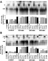
While it is well known that osmotic stimulation induces the expression of Fos family members in the supraoptic nucleus (SON), it is unclear whether the induced protein products are involved in the regulation of the gene transcription of arginine vasopressin (AVP). In the present study, we examined the in vivo correlation between changes in AVP gene transcription and expression of the various Fos family members in the SON after acute osmotic stimuli. The data demonstrated that the peak of AVP transcription (measured by intronic in situ hybridization) observed 15min after an injection of hypertonic saline preceded the expression of Fos proteins, which became detectable at 30min and peaked at 120min. Electrophoretic mobility shift assay showed that the expressed Fos proteins bound to the composite AP-1/CRE-like site in the AVP promoter. These data suggest that Fos proteins in the SON induced by acute osmotic stimuli could affect AVP gene transcription by binding to the AVP promoter, but they are not prerequisite for the induction of AVP gene transcription.
Copyright © 2010 Elsevier Ireland Ltd. All rights reserved.
Figures


Similar articles
-
Expression of immediate early genes and vasopressin heteronuclear RNA in the paraventricular and supraoptic nuclei of rats after acute osmotic stimulus.J Neuroendocrinol. 2005 Apr;17(4):227-37. doi: 10.1111/j.1365-2826.2005.01297.x. J Neuroendocrinol. 2005. PMID: 15842234
-
Intragastric hypertonic saline increases vasopressin and central Fos immunoreactivity in conscious rats.Am J Physiol. 1997 Mar;272(3 Pt 2):R750-8. doi: 10.1152/ajpregu.1997.272.3.R750. Am J Physiol. 1997. PMID: 9087636
-
Robust up-regulation of nuclear red fluorescent-tagged fos marks neuronal activation in green fluorescent vasopressin neurons after osmotic stimulation in a double-transgenic rat.Endocrinology. 2009 Dec;150(12):5633-8. doi: 10.1210/en.2009-0796. Epub 2009 Oct 22. Endocrinology. 2009. PMID: 19850746
-
Fos protein expression in mouse hypothalamic paraventricular (PVN) and supraoptic (SON) nuclei upon osmotic stimulus: colocalization with vasopressin, oxytocin, and tyrosine hydroxylase.Neurochem Int. 2004 Oct;45(5):597-607. doi: 10.1016/j.neuint.2004.04.003. Neurochem Int. 2004. PMID: 15234101
-
Cell-type specific expression of oxytocin and vasopressin genes: an experimental odyssey.J Neuroendocrinol. 2012 Apr;24(4):528-38. doi: 10.1111/j.1365-2826.2011.02236.x. J Neuroendocrinol. 2012. PMID: 21985498 Free PMC article. Review.
Cited by
-
Effects of A-CREB, a dominant negative inhibitor of CREB, on the expression of c-fos and other immediate early genes in the rat SON during hyperosmotic stimulation in vivo.Brain Res. 2012 Jan 6;1429:18-28. doi: 10.1016/j.brainres.2011.10.033. Epub 2011 Oct 26. Brain Res. 2012. PMID: 22079318 Free PMC article.
-
Interactions of the Mechanosensitive Channels with Extracellular Matrix, Integrins, and Cytoskeletal Network in Osmosensation.Front Mol Neurosci. 2017 Apr 5;10:96. doi: 10.3389/fnmol.2017.00096. eCollection 2017. Front Mol Neurosci. 2017. PMID: 28424587 Free PMC article. Review.
-
Transcription factor CREB3L1 mediates cAMP and glucocorticoid regulation of arginine vasopressin gene transcription in the rat hypothalamus.Mol Brain. 2015 Oct 26;8(1):68. doi: 10.1186/s13041-015-0159-1. Mol Brain. 2015. PMID: 26503226 Free PMC article.
References
-
- Arima H, Kondo K, Kakiya S, Nagasaki H, Yokoi H, Yambe Y, Murase T, Iwasaki Y, Oiso Y. Rapid and sensitive vasopressin heteronuclear RNA responses to changes in plasma osmolality. J. Neurondocrinology. 1999;11:337–341. - PubMed
-
- Arima H, Aguilera G. Vasopressin and oxytocin neurones of hypothalamic supraoptic and paraventricular nuclei co-express mRNA for Type-1 and Type-2 corticotropin-releasing hormone receptors. J. Neuroendocrinol. 2000;12:833–842. - PubMed
-
- Arima H, House SB, Gainer H, Aguilera G. Direct stimulation of arginine vasopressin gene transcription by cAMP in parvocellular neurons of the paraventricular nucleus in organotypic cultures. Endocrinology. 2001;142:5027–5030. - PubMed
-
- Arima H, House SB, Gainer H, Aguilera G. Neuronal activity is required for the circadian rhythm of vasopressin gene transcription in the suprachiasmatic nucleus in vivo. Endocrinology. 2002;143:4165–4171. - PubMed
-
- Baler R, Klein D. Circadian expression of transcription factor Fra-2 in the rat pineal gland. J. Biol. Chem. 1995;270:27319–27325. - PubMed
Publication types
MeSH terms
Substances
Grants and funding
LinkOut - more resources
Full Text Sources
Miscellaneous

