

Onchocerca armillata contains the endosymbiotic bacterium Wolbachia and elicits a limited inflammatory response
- PMID: 20850932
- PMCID: PMC3038270
- DOI: 10.1016/j.vetpar.2010.08.031
Onchocerca armillata contains the endosymbiotic bacterium Wolbachia and elicits a limited inflammatory response
Abstract
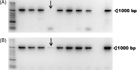
Human onchocerciasis, also known as River Blindness, is a debilitating disease caused by the filarial nematode Onchocerca volvulus. Many, but not all, filarial nematodes carry within their tissues endosymbiotic, Rickettsia-like bacteria of the genus Wolbachia. Onchocerca spp. infections in cattle offer the most relevant, analogous host-parasite model system. West African cattle are commonly co-infected with four Onchocerca spp.; two of these are Wolbachia-positive (Onchocerca gutturosa and Onchocerca ochengi), and the remainder are of unknown Wolbachia status (Onchocerca dukei and Onchocerca armillata). Previous studies have suggested that worm survival is dependent on this bacterium. O. armillata, an abundant parasite of African cattle that has received little attention, is a primitive species that may lack Wolbachia. The objectives of this study were to determine if O. armillata carries Wolbachia and to provide preliminary descriptions of the host inflammatory cell environment around the adult worms. The findings may support or refute the hypothesis that a prime contribution of Wolbachia is to permit long-term survival and reproduction of certain Onchocerca spp. (including O. volvulus in humans). O. armillata adult worms were found in the aorta of 90.7% of cattle (n=54) slaughtered at an abattoir in Ngaoundéré, Adamawa Region, Cameroon. The presence of Wolbachia in O. armillata was confirmed by a specific anti-Wolbachia surface protein antibody detected using a peroxidase conjugate (immunohistochemistry) and PCR for detection of Wolbachia-specific sequences within DNA extracts from frozen worms. Tissue sections stained with haematoxylin and eosin showed the host cell response to be dominated by macrophages and fibroblasts. This is unusual compared with nodule-dwelling Wolbachia-positive Onchocerca spp., where the host response is typically characterised by granulocytes, and suggests that the mechanisms for worm survival employed by this species (which is probably motile) may differ.
Copyright © 2010 Elsevier B.V. All rights reserved.
Figures


References
-
- Alibasoglu M., Golesuk K., Erturk E., Guler S. Türkiyede sigilarde gorulen onchocerciasis olayari (Onchocerca armillata Raillet ve Henry 1909) Vet. Fak. Derg. Ankara Univ. 1969;16:50–60.
-
- al-Zubaidy A.J. Observations on parasitic aortitis in cattle in Iraq. Trans. R. Soc. Trop. Med. Hyg. 1973;67:436. - PubMed
-
- Anon . Final communiqué of the 11th session of the Joint Action Forum (JAF) of APOC. 6 December 2005 (Paris France) APOC; Ouagadougou: 2005.
-
- Atta el Mannan A.M., Hussein H.S., el Sinnary K., Magzoub M. Onchocerca armillata: prevalence and pathology in Sudanese cattle. Ann. Trop. Med. Parasitol. 1984;78:619–625. - PubMed
-
- Bain O., Casiraghi M., Martin C., Uni S. The nematoda Filarioidea: critical analysis linking molecular and traditional approaches. Parasite. 2008;15:342–348. - PubMed
Publication types
MeSH terms
Grants and funding
LinkOut - more resources
Full Text Sources
Miscellaneous
