. 2010 Nov 5;403(4):616-29.
doi: 10.1016/j.jmb.2010.09.022. Epub 2010 Sep 17.

Structure-based analysis of Toxoplasma gondii profilin: a parasite-specific motif is required for recognition by Toll-like receptor 11

Affiliations

Structure-based analysis of Toxoplasma gondii profilin: a parasite-specific motif is required for recognition by Toll-like receptor 11

Kaury Kucera et al. J Mol Biol. .

Abstract

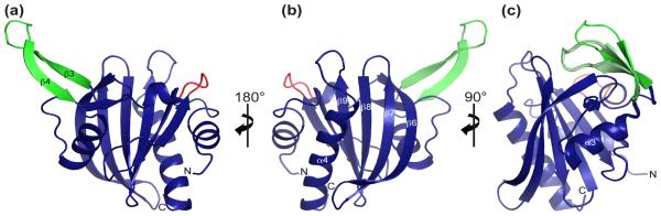
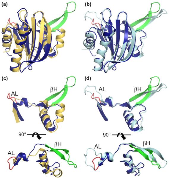
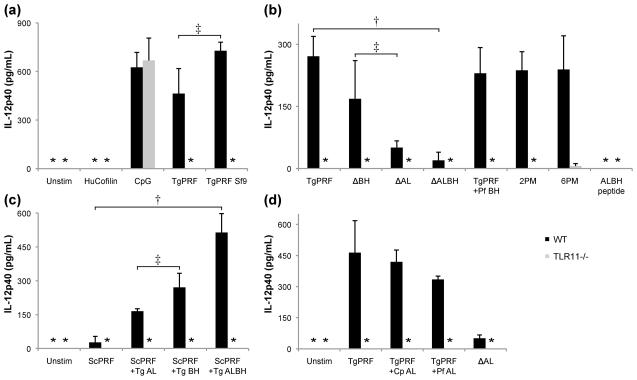
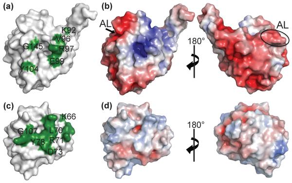
Profilins promote actin polymerization by exchanging ADP for ATP on monomeric actin and delivering ATP-actin to growing filament barbed ends. Apicomplexan protozoa such as Toxoplasma gondii invade host cells using an actin-dependent gliding motility. Toll-like receptor (TLR) 11 generates an innate immune response upon sensing T. gondii profilin (TgPRF). The crystal structure of TgPRF reveals a parasite-specific surface motif consisting of an acidic loop, followed by a long β-hairpin. A series of structure-based profilin mutants show that TLR11 recognition of the acidic loop is responsible for most of the interleukin (IL)-12 secretion response to TgPRF in peritoneal macrophages. Deletion of both the acidic loop and the β-hairpin completely abrogates IL-12 secretion. Insertion of the T. gondii acidic loop and β-hairpin into yeast profilin is sufficient to generate TLR11-dependent signaling. Substitution of the acidic loop in TgPRF with the homologous loop from the apicomplexan parasite Cryptosporidium parvum does not affect TLR11-dependent IL-12 secretion, while substitution with the acidic loop from Plasmodium falciparum results in reduced but significant IL-12 secretion. We conclude that the parasite-specific motif in TgPRF is the key molecular pattern recognized by TLR11. Unlike other profilins, TgPRF slows nucleotide exchange on monomeric rabbit actin and binds rabbit actin weakly. The putative TgPRF actin-binding surface includes the β-hairpin and diverges widely from the actin-binding surfaces of vertebrate profilins.


Figures

Fig. 1
Fig. 1. Overall structure of Toxoplasma gondii profilin (TgPRF)
(a-c) Ribbon representations of TgPRF in blue with the acidic loop and β-hairpin highlighted in red and green respectively. (b) TgPRF is in the standard profilin orientation, which shows the putative actin binding surface. (c) An overlay of all four molecules in the asymmetric unit using residues 38-80 as the reference shows flexibility in the extended β-hairpin. β-hairpin residues 50-67 have an average RMSD of 0.50 Å. The average main chain RMSD between the four molecules is 0.15 Å.
Fig. 2
Fig. 2. Sequence alignment of apicomplexan protozoan profilins
Secondary structure is shown for T. gondii profilin (TgPRF) on the top, and for S. cerevisiae, representing the conserved non-apicomplexan profilin structure, on the bottom. Secondary structure nomenclature follows the canonical profilin fold. Strictly conserved residues are highlighted in black, orange denotes conserved residues and in yellow are residues absolutely conserved in the parasites most closely related to TgPRF. The apicomplexan-specific acidic loop (AL) and β-hairpin (βH) are boxed in red and green, respectively. GenBank accession numbers are: T. gondii 61612092, Cryptosporidium parvum 126644761, Cryptosporidium hominis 67593937, Eimeria acervulina 405637, Eimeria tenella 117960055, Plasmodium knowlesi 193808670, Plasmodium falciparum 206581653, Theileria annulata 84994870, Theileria parva 71030962, Babesia bovis 78458472, Saccharomyces cerevisiae 6324696. The sequence for Neospora caninum is from toxoDB, accession no. NC_LIV_10440.
Fig. 3
Fig. 3. Structural comparison of profilins from T. gondii, S. cerevisiae and P. falciparum
T. gondii profilin (TgPRF) is in blue with red acidic loop and green β-hairpin. (a) Superposition of TgPRF onto S. cerevisiae profilin (PDB code 1YPR) in orange, which represents the conserved non-apicomplexan profilin structure (RMSD is 2.35 Å). (b) Superposition of TgPRF onto P. falciparum profilin (PfPRF, PDB code 2JKF) in cyan (RMSD is 1.06 Å). (c, d) The divergent features of TgPRF include a highly acidic loop (AL, TgPRF residues 37-40), and a β-hairpin (βH, residues 50-67). The latter is conserved among apicomplexans in length and overall structure, but not in sequence.
Fig. 4
Fig. 4. TLR11 recognizes specific features of TgPRF
(a) Peritoneal macrophages from wild-type (WT) and TLR11−/−, mice were stimulated for 24 h with 3.0 μg/ml of either TgPRF, TgPRF purified from Sf9 insect cells, or 5 mM of CpG as a positive control. Macrophages were stimulated with 3.0 μg/ml of human cofilin as a negative control. (b) Peritoneal macrophages were stimulated with 3.0 μg/ml of either TgPRF, acidic loop deletion mutant TgPRF (ΔAL), β-hairpin deletion mutant TgPRF (ΔBH), double deletion mutant TgPRF (ΔALBH), a TgPRF mutant with P. falciparum β-hairpin (TgPRF +Pf BH), actin-binding surface TgPRF point mutants 2PM or 6PM and a peptide with the acidic loop and β-hairpin sequence (ALBH peptide). (c) Peritoneal macrophages were stimulated with TgPRF, a TgPRF mutant with the acidic loop from C. parvum (TgPRF +Cp AL), a TgPRF mutant with the acidic loop from P. falciparum (TgPRF +Pf AL) and a TgPRF acidic loop deletion mutant that, like S. cerevisiae profilin, has two glycines in place of the acidic loop (TgPRF +Sc AL). (d) Peritoneal macrophages were stimulated with S. cerevisiae profilin (ScPRF), a ScPRF mutant containing the T. gondii acidic loop (ScPRF +Tg AL), a ScPRF mutant containing the T. gondii β-hairpin (ScPRF +Tg BH), and a ScPRF mutant containing both the acidic loop and β-hairpin (ScPRF +Tg ALBH). After 24 h, supernatant was removed and analyzed for IL-12p40 levels by ELISA. Each bar represents the mean ± SD of triplicate measurements of three independent experiments. Statistical analysis was performed using the Student's t test where † indicates P < 0.01; ‡ indicates P > 0.05 arbitrary units. In the columns marked with an asterisk, the IL-12p40 concentration was below detectable levels. Together these data show that the parasite-specific motif in TgPRF is the key determinant for recognition by TLR11, with the acidic loop as the primary determinant and the β-hairpin as a secondary but necessary determinant.
Fig. 5
Fig. 5. Effect of TgPRF on the rate of nucleotide exchange of ATP bound monomeric actin
(a) Time courses of ε-ATP exchange from actin monomers in the absence (red trace) and presence of TgPRF (black traces; 10–60 μM from left to right. The solid lines through the data represent the best fits to single exponentials. The final concentrations were 0.5 μM monomeric actin, 2.5 μM εATP, 2 mM ATP, 10–60 μM TgPRF. Shown for comparison are the effects of 1.7 μM human profilin (HuPfn, blue trace) and 1.1 μM cofilin (green trace). The inset shows the same data and fits over shorter timescale for visualization. (b) TgPRF concentration-dependence of the observed ATP exchange rate constant (black). The ε-ATP exchange rate constant of actin alone varied from 0.03 to 0.1 s−1 for various preparations. For comparison are shown: TgPRF β-hairpin deletion mutant (ΔBH, orange), TgPRF acidic loop deletion mutant (ΔAL, pink), TgPRF point mutant 6PM (brown), and human cofilin (green). The lines represent the best fits of the data to Equation 1 (see Materials and Methods), which yields Kp = 13.9 ± 5.0 μM for wild-type TgPRF.
Fig. 6
Fig. 6. Surface representations of profilins from T. gondii and S. cerevisiae
Comparison of the surface representations of Toxoplasma gondii profilin TgPRF (top row) to S. cerevisiae profilin (bottom row) suggests divergent profilin function. (a) TgPRF residues that are conserved with known actin-binding residues in S. cerevisiae, S. pombe and B. taurus profilins are shown in green and labeled. (b) S. cerevisiae profilin residues known to bind actin monomers are shown in green. Only residues that share conservation with putative TgPRF actin binding residues are labeled. (c) Comparison of electrostatic surface charge shows polarization of the TgPRF surface. (d) In comparison, the surface of S. cerevisiae is more uniformly neutral. Positive and negative electrostatic protein surface potentials, contoured from +5 kT to −5 kT, are shown in blue and red respectively. The TgPRF acidic loop (AL) is labeled for reference.

References

    1. Montoya JG, Liesenfeld O. Toxoplasmosis. Lancet. 2004;363:1965–1976. - PubMed
    1. Dobrowolski JM, Sibley LD. Toxoplasma invasion of mammalian cells is powered by the actin cytoskeleton of the parasite. Cell. 1996;84:933–939. - PubMed
    1. Sahoo N, Beatty W, Heuser J, Sept D, Sibley LD. Unusual kinetic and structural properties control rapid assembly and turnover of actin in the parasite Toxoplasma gondii. Mol Biol Cell. 2006;17:895–906. - PMC - PubMed
    1. Schmitz S, Grainger M, Howell S, Calder LJ, Gaeb M, Pinder JC, Holder AA, Veigel C. Malaria parasite actin filaments are very short. J Mol Biol. 2005;349:113–125. - PubMed
    1. Schuler H, Mueller AK, Matuschewski K. Unusual properties of Plasmodium falciparum actin: new insights into microfilament dynamics of apicomplexan parasites. FEBS Lett. 2005;579:655–660. - PubMed

Publication types

MeSH terms

Associated data

LinkOut - more resources