. 2010 Dec 3;486(1):47-50.
doi: 10.1016/j.neulet.2010.09.042. Epub 2010 Sep 17.

Neuromelanin inhibits CXCL10 expression in human astroglial cells

Affiliations

Neuromelanin inhibits CXCL10 expression in human astroglial cells

Neda Saffarian Tousi et al. Neurosci Lett. .

Abstract

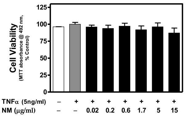
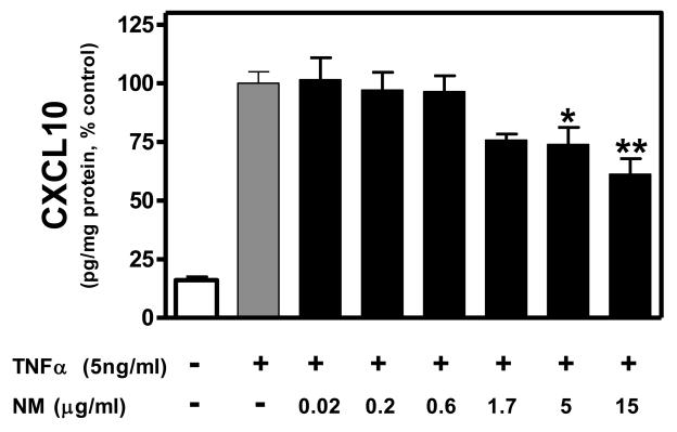
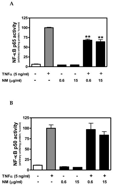
Increasing evidence indicates neuroinflammation is instrumental in the pathogenesis of Parkinson's disease (PD). In PD, there is selective degeneration of neuromelanin (NM)-containing dopamine neurons. Neuromelanin is predominantly cytoprotective within dopaminergic neurons, whereas, NM released from damaged neurons activates microglia. However, the effects of NM on astroglial cells remain largely unknown. Astroglia are essential to neuronal homeostasis and responsive to injury, in part, through secretion of chemokines, including interferon γ inducible protein-10 (CXCL10). Thus, we used an in vitro approach to identify the effects of NM on TNFα-induced CXCL10 expression in human astroglial cells. TNFα-induced CXCL10 expression was inhibited in NM exposed cells. Additionally, TNFα-induced NF-кB activation was inhibited by NM. Given that CXCL10 expression is NF-кB-dependent in human astroglial cells, these findings suggest that NM may inhibit CXCL10 expression, in part, through an NF-кB-dependent mechanism. While the in vivo consequences of NM mediated effects on astroglial CXCL10 expression remain to be fully elucidated, insights obtained in this study further our understanding of the effects of NM on inflammatory signaling in human astroglial cells.


Figures

Figure 1
Figure 1. Neuromelanin is not cytotoxic to TNFα-stimulated human A172 astroglial cells
A172 cells were cultured in serum free medium for 48 h in the presence or absence of NM (0.02-15 μg/ml), then co-exposed to human recombinant TNFα (5ng/ml) for an additional 24 h. To assess mitochondrial integrity/cell viability, the MTT assay was performed. Data were normalized as percent control (TNFα alone) within each experiment and presented as the mean + S.E.M. of triplicate measures from 2-3 independent experiments. No significant differences (P < 0.05) among treatments were detected by one-way ANOVA.
Figure 2
Figure 2. Neuromelanin inhibits TNFα-induced CXCL10 protein expression in human A172 astroglial cells
A172 cells were cultured in serum free medium for 48 h in the presence or absence of NM (0.02-15 μg/ml), then co-exposed to human recombinant TNFα (5ng/ml) for an additional 24 h. Secreted CXCL10 protein in the culture media was then quantitated using a standard dual-antibody solid phase immunoassay, according to the manufacturer's instructions as previously described [12]. Absorbance was read at 450 nm (with wavelength correction set at 650 nm) on a BIOTEK HT spectrophotometer. Data were normalized as percent control (TNFα alone) within each experiment and presented as the mean + S.E.M. of triplicate measures from 3-4 independent experiments. Statistical differences determined by one-way ANOVA with Dunnett's post-hoc comparisons. *P < 0.05 vs TNFα; **P < 0.01 vs TNFα.
Figure 3
Figure 3. Neuromelanin inhibits TNFα-induced NF-κB activation in human A172 astroglial cells
A172 cells were cultured in serum free medium for 72 h in the presence or absence of NM (0.6 or 15 μg/ml). Cells were co-exposed to human recombinant TNFα (5ng/ml) for the final 0.5 h of the 72 h NM exposure period. Nuclear protein fractions were obtained and NF-κB p65- and NF-κB p50-DNA binding activity was determined using a Transcription Factor kit, according to manufacturer's instructions. Binding activity was determined by luminescence of the labeled NF-κB-DNA product as measured using a BIO-TEK HT spectrophotometer. Binding activity was normalized to total nuclear protein and presented as arbitrary units. Data represent the mean + S.E.M. of duplicate measures from 2 independent experiments. Significant differences were determined by one-way ANOVA with Dunnett's post-hoc comparisons. **P < 0.01 vs TNFα.

References

    1. Agrawal L, Louboutin JP, Marusich E, Reyes BA, Van Bockstaele EJ, Strayer DS. Dopaminergic neurotoxicity of HIV-1 gp120: reactive oxygen species as signaling intermediates. Brain Res. 2010;1306:116–130. - PubMed
    1. Aoki E, Yano R, Yokoyama H, Kato H, Araki T. Role of nuclear transcription factor kappa B (NF-kappaB) for MPTP (1-methyl-4-phenyl-1,2,3,6-tetrahyropyridine)-induced apoptosis in nigral neurons of mice. Exp Mol Pathol. 2009;86:57–64. - PubMed
    1. Babcock AA, Kuziel WA, Rivest S, Owens T. Chemokine expression by glial cells directs leukocytes to sites of axonal injury in the CNS. J Neurosci. 2003;23:7922–7930. - PMC - PubMed
    1. Banati RB, Daniel SE, Blunt SB. Glial pathology but absence of apoptotic nigral neurons in long-standing Parkinson's disease. Mov Disord. 1998;13:221–227. - PubMed
    1. Beach TG, Sue LI, Walker DG, Lue LF, Connor DJ, Caviness JN, Sabbagh MN, Adler CH. Marked microglial reaction in normal aging human substantia nigra: correlation with extraneuronal neuromelanin pigment deposits. Acta Neuropathol. 2007;114:419–424. - PubMed

Publication types

MeSH terms

LinkOut - more resources