

Challenges to attention: a continuous arterial spin labeling (ASL) study of the effects of distraction on sustained attention
- PMID: 20851189
- PMCID: PMC2997179
- DOI: 10.1016/j.neuroimage.2010.09.026
Challenges to attention: a continuous arterial spin labeling (ASL) study of the effects of distraction on sustained attention
Abstract
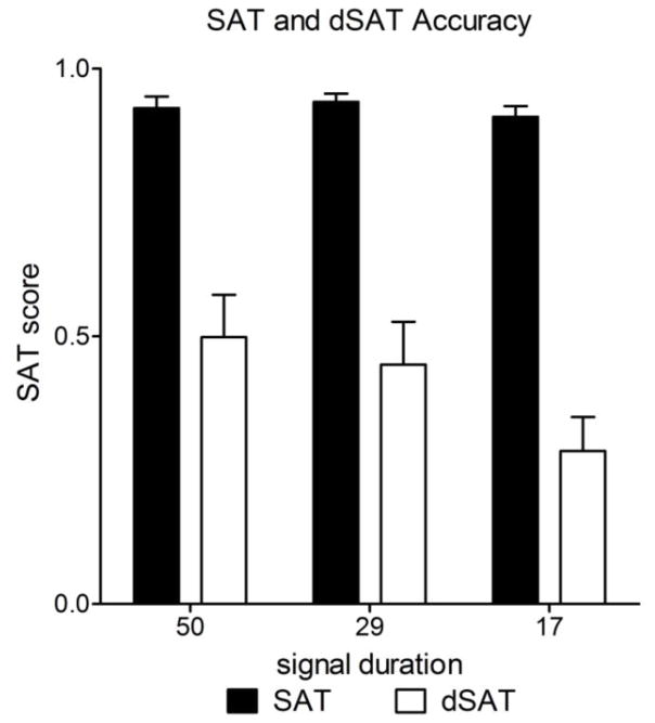
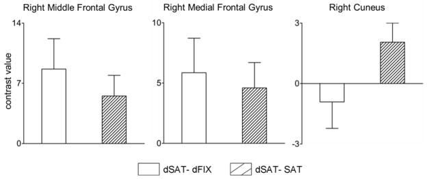
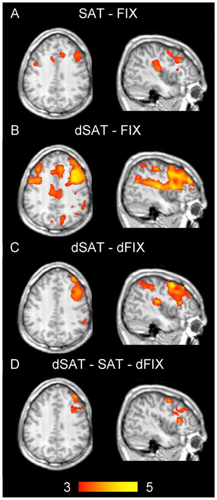
Maintaining attention and performance over time is an essential part of many activities, and effortful cognitive control is required to avoid vigilance decrements and interference from distraction. Regions at or near right middle frontal gyrus (Brodmann's area (BA) 9), as well as in other prefrontal and parietal areas, are often activated in studies of sustained attention (e.g., Cabeza and Nyberg, 2000; Kim et al., 2006; Lim et al., 2010). This activation has often been interpreted as representing the engagement of cognitive control processes. However, such studies are typically implemented at one level of task difficulty, without an experimental manipulation of control demands. The present study used the distractor condition sustained attention task (dSAT), which has been used extensively in animals to determine the role of neuromodulator systems in attentional performance, to test the hypotheses that BA 9 is sensitive to changes in the demand for cognitive control and that this sensitivity reflects an increased engagement of attentional effort. Continuous arterial spin labeling (ASL) was used to measure neural activity in sixteen healthy, young adults performing a sustained attention task under standard conditions and under a distraction condition that provided an experimental manipulation of demands on cognitive control. The distractor impaired behavioral performance and increased activation in right middle frontal gyrus. Larger increases in right middle frontal gyrus activity were associated with greater behavioral vulnerability to the distractor. These findings indicate that while right middle frontal gyrus regions are sensitive to demands for attentional effort and control, they may not be sufficient to maintain performance under challenge. In addition, they demonstrate the sensitivity of ASL methods to variations in task demands, and suggest that the dSAT may be a useful tool for translational cross-species and clinical research.
Copyright © 2010 Elsevier Inc. All rights reserved.
Figures


Similar articles
-
Continuous ASL perfusion fMRI investigation of higher cognition: quantification of tonic CBF changes during sustained attention and working memory tasks.Neuroimage. 2006 May 15;31(1):376-85. doi: 10.1016/j.neuroimage.2005.11.035. Epub 2006 Jan 19. Neuroimage. 2006. PMID: 16427324 Free PMC article.
-
Distinct Frontoparietal Networks Underlying Attentional Effort and Cognitive Control.J Cogn Neurosci. 2017 Jul;29(7):1212-1225. doi: 10.1162/jocn_a_01112. Epub 2017 Mar 2. J Cogn Neurosci. 2017. PMID: 28253080 Free PMC article.
-
Probing the Neural Mechanisms for Distractor Filtering and Their History-Contingent Modulation by Means of TMS.J Neurosci. 2019 Sep 18;39(38):7591-7603. doi: 10.1523/JNEUROSCI.2740-18.2019. Epub 2019 Aug 6. J Neurosci. 2019. PMID: 31387915 Free PMC article.
-
γ-Aminobutyric acid type A receptor binding affinity in the right inferior frontal gyrus at resting state predicts the performance of healthy elderly people in the visual sustained attention test.Int Psychogeriatr. 2018 Sep;30(9):1385-1391. doi: 10.1017/S1041610217002988. Epub 2018 Mar 21. Int Psychogeriatr. 2018. PMID: 29559018
-
The neural substrates associated with attentional resources and difficulty of concurrent processing of the two verbal tasks.Neuropsychologia. 2012 Jul;50(8):1998-2009. doi: 10.1016/j.neuropsychologia.2012.04.025. Epub 2012 May 6. Neuropsychologia. 2012. PMID: 22571931
Cited by
-
Transient Distraction and Attentional Control during a Sustained Selective Attention Task.J Cogn Neurosci. 2016 Jul;28(7):935-47. doi: 10.1162/jocn_a_00949. Epub 2016 Mar 11. J Cogn Neurosci. 2016. PMID: 26967946 Free PMC article.
-
The presynaptic choline transporter imposes limits on sustained cortical acetylcholine release and attention.J Neurosci. 2013 Feb 6;33(6):2326-37. doi: 10.1523/JNEUROSCI.4993-12.2013. J Neurosci. 2013. PMID: 23392663 Free PMC article.
-
The functional oculomotor network and saccadic cognitive control in healthy elders.Neuroimage. 2014 Jul 15;95:61-8. doi: 10.1016/j.neuroimage.2014.03.051. Epub 2014 Mar 24. Neuroimage. 2014. PMID: 24675647 Free PMC article.
-
Developing treatments for cognitive deficits in schizophrenia: the challenge of translation.J Psychopharmacol. 2015 Feb;29(2):178-96. doi: 10.1177/0269881114555252. Epub 2014 Dec 16. J Psychopharmacol. 2015. PMID: 25516372 Free PMC article. Review.
-
Deficits in prefrontal metabotropic glutamate receptor 5 are associated with functional alterations during emotional processing in bipolar disorder.J Affect Disord. 2024 Sep 15;361:415-424. doi: 10.1016/j.jad.2024.06.025. Epub 2024 Jun 13. J Affect Disord. 2024. PMID: 38876317
References
-
- Aguirre G, Detre J, et al. Experimental design and the relative sensitivity of BOLD and perfusion fMRI. Neuroimage. 2002;13:488–500. - PubMed
-
- Arnold HM, Burk JA, Hodgson EM, Sarter M, Bruno JP. Differential cortical acetylcholine release in rats performing a sustained attention task versus behavioral control tasks that do not explicitly tax attention. Neuroscience. 2002;114:451–460. - PubMed
-
- Baddeley AD. Working Memory. Oxford University Press; Oxford: 1986.
-
- Bakeman R. Recommended effect size statistics for repeated measures designs. Behav Res Methods. 2005;37:379–384. - PubMed
-
- Beckmann CF, Jenkinson M, Smith SM. General multilevel linear modeling for group analysis in FMRI. Neuroimage. 2003;20:1052–1063. - PubMed
Publication types
MeSH terms
Substances
Grants and funding
LinkOut - more resources
Full Text Sources
