. 2010 Sep 24;17(9):1008-17.
doi: 10.1016/j.chembiol.2010.06.016.

The quaternary organization and dynamics of the molecular chaperone HSP26 are thermally regulated

Affiliations

The quaternary organization and dynamics of the molecular chaperone HSP26 are thermally regulated

Justin L P Benesch et al. Chem Biol. .

Abstract

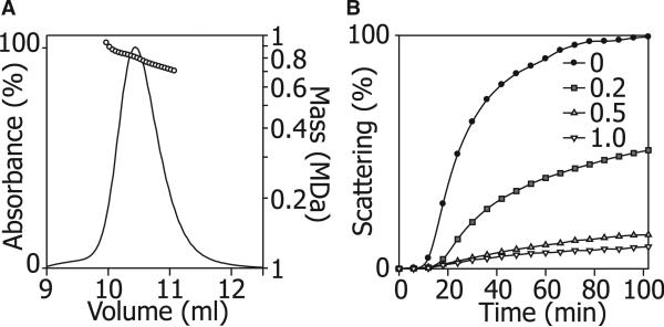
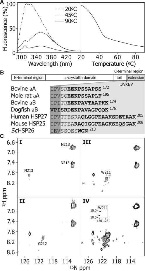
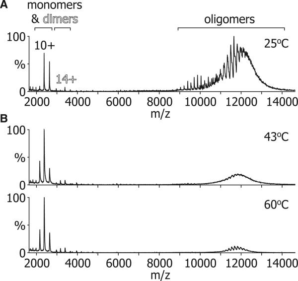
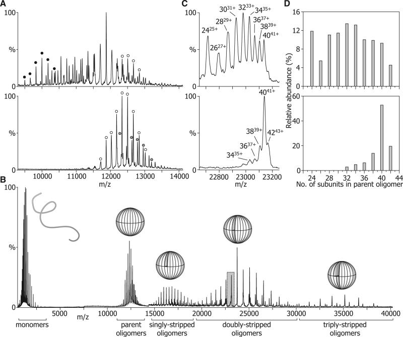
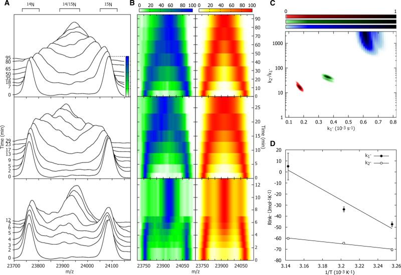
The function of ScHSP26 is thermally controlled: the heat shock that causes the destabilization of target proteins leads to its activation as a molecular chaperone. We investigate the structural and dynamical properties of ScHSP26 oligomers through a combination of multiangle light scattering, fluorescence spectroscopy, NMR spectroscopy, and mass spectrometry. We show that ScHSP26 exists as a heterogeneous oligomeric ensemble at room temperature. At heat-shock temperatures, two shifts in equilibria are observed: toward dissociation and to larger oligomers. We examine the quaternary dynamics of these oligomers by investigating the rate of exchange of subunits between them and find that this not only increases with temperature but proceeds via two separate processes. This is consistent with a conformational change of the oligomers at elevated temperatures which regulates the disassembly rates of this thermally activated protein.


Figures

Figure 1
Figure 1. Oligomeric Distribution and Chaperone Activity of ScHSP26
(A) ScHSP26 examined by SEC-MALS illustrates the heterogeneity of the oligomeric ensemble. The molecular mass decreased across the peak from 950 kDa to 580 kDa and was centered around a maximum population species of 810 kDa, corresponding to an approximately 34 subunit oligomer. (B) Insulin reduction assay of the chaperone action of ScHSP26. At 25°C, in the absence of ScHSP26, insulin B chain is reduced and exhibits an apparent increase in absorbance at 360 nm due to light scattering over a period of 100 min as a result of aggregation. At a subunit molar ratio of 0.2:1.0 ScHSP26:insulin, a 50% reduction in aggregation was observed due to the chaperone action of ScHSP26. At the higher ratios of 0.5:1.0 and 1.0:1.0, almost complete suppression of aggregation is achieved.
Figure 2
Figure 2. C-Terminal Flexibility of ScHSP26
(A) The dependence of tertiary/quaternary structure of the ScHSP26 oligomers on temperature was examined by observing the changes in intrinsic tryptophan fluorescence. ScHSP26 was heated from 20–90°C at 1°C/min (right). The sample was excited at 280 nm and emission was recorded at 330 nm. The resultant melt demonstrated that the tryptophan residues became significantly more solvent exposed between 20°C and 45°C, whereas a much smaller, linear decrease in fluorescence was observed up to 90°C. Fluorescence spectra were also recorded at three temperatures to provide information on the structural transitions (left). At 20°C, the emission spectrum has a λmax at ≈325 nm. At 45°C, there was a decrease in the emission intensity concomitant with a dramatic shift in the λmax to 352 nm. At 90°C, a further decrease in fluorescence intensity but no λmax shift occurred. These data indicate that the majority of the tertiary structure in the vicinity of the tryptophan residues is lost during the initial unfolding transition. (B) The primary sequence of sHSPs in general is composed of an N-terminal region, the α-crystallin domain, and a C-terminal region. The latter is subdivided into a C-terminal tail, and a C-terminal extension, separated by the conserved I/VXI/V motif. The sequence of ScHSP26 is aligned to those of animal sHSPs which have been demonstrated to contain flexible C-terminal extensions. Residues which are clearly identified by NMR spectroscopy measurements are highlighted in bold. (C) 1H-15N 2D HSQC NMR spectra of ScHSP26 at I: 25°C and 1.66 mM; II: 45°C and 1.66 mM; III: 45°C and 0.33 mM; IV: 45°C and 0.17 mM. At the same concentration (1.66 mM), solutions of HSP26 exhibit a minor enhancement of C-terminal flexibility with increased temperature, as evidenced by the greater number of cross-peaks resolved at random coil chemical shift values and an additional cross-peak from G212 (I and II). At 45°C, 5- and 10-fold dilution an increased number of cross-peaks are observed (III and IV), suggesting greater C-terminal flexibility. This is consistent with an overall loosening of the structure and smaller, less-structured species of ScHSP26 being present under more dilute conditions.
Figure 3
Figure 3. MS Demonstrates a Temperature-Dependent Dissociation of ScHSP26
(A) At 25°C the mass spectrum of ScHSP26 was dominated by signal arising from a broad range of oligomers with a minor percentage of monomer and dimer. (B) At higher temperatures (43°C and 60°C), the relative amount of signal arising from the ScHSP26 oligomers was greatly diminished with a concomitant increase in the percentage of monomer and dimer.
Figure 4
Figure 4. Defining the Oligomeric Distributions of ScHSP26
(A) NanoESI-MS demonstrated that ScHSP26 exists as a broad range of oligomers at 25°C (upper), but forms mainly 40 mers (open circles), and 42 mers (shaded circles) at 43°C (lower). Note that a 24-mer is notably abundant at room temperature (black circles). (B) Collision-induced dissociation of monomers from the native oligomers gives rise to a series of stripped oligomers of reduced charge with sufficient peak resolution to accurately define their size distribution. Overlaid are schematic representations of the different species. (C) Portion of the doubly-stripped region highlighted in (B) for ScHSP26 at 25°C (upper) and 43°C (lower). From the peaks in this region of the spectra, it was possible to assign charge states to individual oligomers, as well as calculate their relative abundance. (D) Histograms showing the most abundant oligomers present at 25°C and 43°C after analysis of the stripped oligomer region.
Figure 5
Figure 5. Subunit Exchange
(A) The overlapping peaks of the doubly stripped oligomers were monitored at 34°C (top), 39°C (middle), and 45°C (bottom) until exchange was complete. 14N and 15N-labeled ScHSP26 homo-oligomer peaks diminished with time, and in their place a peak corresponding to hetero-oligomers of the two subunits arose. This peak was broad because of the variety of isotopic stoichiometries in the polydisperse assembly. As expected, the rate of exchange was found to increase with temperature; notably, however, there appeared to be a change in the mechanism of exchange at 45°C. This was evidenced by the fact that in the period between 2 and 4 min a population of fully exchanged ScHSP26 coexisted with the original homo-oligomers. (B) Alternative “top down” contour plot representation of the data in (A), left, compared with the simulated subunit exchange time-course that best fits each temperature, right. (C) Reduced χ2 surface for the fitting of subunit exchange data to two rate constants, k1 and k2. (D) Arrhenius plots for the temperature dependence of k1 and k2 allows the extraction of the activation energies of the two processes. Mean values and error estimates are obtained by fitting a two-dimensional Gaussian function of the form a*exp(–(xx0)2/(2cx2))*exp(–(yy0)2/(2cy2)) to the χ2 probability surface. The central position of the Gaussian function indicates the mean value of the fitting parameter (x0, y0), and the widths the uncertainties (cx, cy).

References

    1. Aquilina JA, Benesch JLP, Bateman OA, Slingsby C, Robinson CV. Polydispersity of a mammalian chaperone: mass spectrometry reveals the population of oligomers in alphaB-crystallin. Proc. Natl. Acad. Sci. USA. 2003;100:10611–10616. - PMC - PubMed
    1. Aquilina JA, Benesch JLP, Ding LL, Yaron O, Horwitz J, Robinson CV. Subunit exchange of polydisperse proteins: mass spectrometry reveals consequences of alphaA-crystallin truncation. J. Biol. Chem. 2005;280:14485–14491. - PubMed
    1. Basha E, Jones C, Wysocki V, Vierling E. Mechanistic differences between two conserved classes of small heat shock proteins found in the plant cytosol. J. Biol. Chem. 2010;285:11489–11497. - PMC - PubMed
    1. Basha E, Lee GJ, Demeler B, Vierling E. Chaperone activity of cytosolic small heat shock proteins from wheat. Eur. J. Biochem. 2004;271:1426–1436. - PubMed
    1. Benesch JLP. Collisional activation of protein complexes: picking up the pieces. J. Am. Soc. Mass Spectrom. 2009;20:341–348. - PubMed

Publication types

MeSH terms

Substances

LinkOut - more resources