. 2011 May;21(5):1066-75.
doi: 10.1093/cercor/bhq180. Epub 2010 Sep 17.

The neural organization of semantic control: TMS evidence for a distributed network in left inferior frontal and posterior middle temporal gyrus

Affiliations

The neural organization of semantic control: TMS evidence for a distributed network in left inferior frontal and posterior middle temporal gyrus

Carin Whitney et al. Cereb Cortex. 2011 May.

Abstract

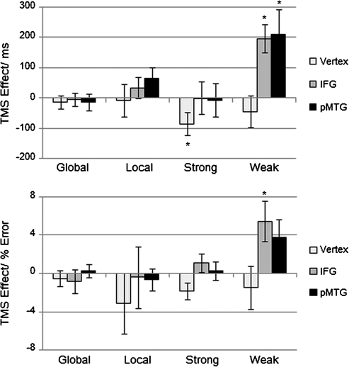
Assigning meaning to words, sounds, and objects requires stored conceptual knowledge plus executive mechanisms that shape semantic retrieval according to the task or context. Despite the essential role of control in semantic cognition, its neural basis remains unclear. Neuroimaging and patient research has emphasized the importance of left inferior frontal gyrus (IFG)--however, impaired semantic control can also follow left temporoparietal lesions, suggesting that this function may be underpinned by a large-scale cortical network. We used repetitive transcranial magnetic stimulation in healthy volunteers to disrupt processing within 2 potential sites in this network--IFG and posterior middle temporal cortex. Stimulation of both sites selectively disrupted executively demanding semantic judgments: semantic decisions based on strong automatic associations were unaffected. Performance was also unchanged in nonsemantic tasks--irrespective of their executive demands--and following stimulation of a control site. These results reveal that an extended network of prefrontal and posterior temporal regions underpins semantic control.


Figures

Figure 1.
Figure 1.
Semantic control in the brain. Overlap image of TMS stimulation sites (“orange”), lesions in patients with deregulated semantic control after infarction to prefrontal and/or temporoparietal cortex (“red”), and brain activation for high > low executive semantic demands during fMRI (“black,” “green,” “purple”). Activation peaks correspond to studies that were used to generate coordinates of stimulation sites. Note: black = Wagner et al. (2001), green = Badre et al. (2005), and purple = Thompson-Schill et al. (1997).
Figure 2.
Figure 2.
Stimuli. Example trials for the semantic (“left panel”) and nonsemantic tasks (“right panel”). Participants had to select the target word that was either strongly related to the cue shown above (strong association) or weakly related (weak association). In the Navon tasks, participants had to choose the target compound letter that resembles the cue letter either in its global shape (global Navon) or in its local, smaller elements (local Navon). Note: Target items are underlined, and Navon compound letters are enlarged for illustration purposes.
Figure 3.
Figure 3.
Stimulation sites. rTMS was delivered to the pars triangularis of left IFG, the left pMTG, and the vertex (not shown). The image on the right depicts probability maps for BA 44 and 45, available from the SPM Anatomy toolbox. Note: Coordinates are in MNI space; orange = pars triangularis, yellow = inferior and superior temporal sulcus, purple = Sylvian fissure.
Figure 4.
Figure 4.
Behavioral results. Reaction time (RT) and error rate at baseline (no TMS) and directly after stimulation (TMS) to the vertex, left IFG, and pMTG. Note: global = global Navon task, local = local Navon task, strong = strong cue–target association strength, weak = weak cue–target association strength.
Figure 5.
Figure 5.
TMS effects. Difference in reaction time (top) and error rate (bottom) between TMS and baseline performance (TMS – no TMS) for each stimulation site. Positive values indicate a decline in performance after brain stimulation, while negative values indicate improvement. Note: global = global Navon task, local = local Navon task, strong = strong cue–target association strength, weak = weak cue–target association strength. * P < 0.05.

Similar articles

Cited by

References

    1. Badre D. Cognitive control, hierarchy, and the rostro-caudal organization of the frontal lobes. Trends Cogn Sci. 2008;12:193–200. - PubMed
    1. Badre D, D'Esposito M. Functional magnetic resonance imaging evidence for a hierarchical organization of the prefrontal cortex. J Cogn Neurosci. 2007;19:2082–2099. - PubMed
    1. Badre D, Poldrack RA, Pare-Blagoev EJ, Insler RZ, Wagner AD. Dissociable controlled retrieval and generalized selection mechanisms in ventrolateral prefrontal cortex. Neuron. 2005;47:907–918. - PubMed
    1. Badre D, Wagner AD. Left ventrolateral prefrontal cortex and the cognitive control of memory. Neuropsychologia. 2007;45:2883–2901. - PubMed
    1. Bedny M, McGill M, Thompson-Schill SL. Semantic adaptation and competition during word comprehension. Cereb Cortex. 2008;18:2574–2585. - PMC - PubMed

Publication types