. 2010 Dec;95(12):2063-71.
doi: 10.3324/haematol.2010.025783. Epub 2010 Sep 17.

HuMab-7D8, a monoclonal antibody directed against the membrane-proximal small loop epitope of CD20 can effectively eliminate CD20 low expressing tumor cells that resist rituximab-mediated lysis

Affiliations

HuMab-7D8, a monoclonal antibody directed against the membrane-proximal small loop epitope of CD20 can effectively eliminate CD20 low expressing tumor cells that resist rituximab-mediated lysis

Tom van Meerten et al. Haematologica. 2010 Dec.

Abstract

Background: Incorporation of the chimeric CD20 monoclonal antibody rituximab in the treatment schedule of patients with non-Hodgkin's lymphoma has significantly improved outcome. Despite this success, about half of the patients do not respond to treatment or suffer from a relapse and additional therapy is required. A low CD20-expression level may in part be responsible for resistance against rituximab. We therefore investigated whether the CD20-expression level related resistance to rituximab could be overcome by a new group of CD20 mAbs (HuMab-7D8 and ofatumumab) targeting a unique membrane-proximal epitope on the CD20 molecule.

Design and methods: By retroviral transduction of the CD20 gene into CD20-negative cells and clonal selection of transduced cells a system was developed in which the CD20-expression level is the only variable. These CD20 transduced cells were used to study the impact of rituximab and HuMab-7D8 mediated complement-dependent cytotoxicity. To study the in vivo efficacy of these mAbs an in vivo imaging system was generated by retroviral expression of the luciferase gene in the CD20-positive cells.

Results: We show that HuMab-7D8 efficiently killed CD20(low) cells that are not susceptible to rituximab-induced killing in vitro. In a mouse xenograft model, we observed a comparable increase in survival time between HuMab-7D8 and rituximab-treated mice. Most significantly, however, HuMab-7D8 eradicated all CD20-expressing cells both in the periphery as well as in the bone marrow whereas after rituximab treatment CD20(low) cells survived.

Conclusions: Cells that are insensitive to in vitro and in vivo killing by rituximab as the result of their low CD20-expression profile may be efficiently killed by an antibody against the membrane-proximal epitope on CD20. Such antibodies should, therefore, be explored to overcome rituximab resistance in the clinic.


Figures

Figure 1.
Figure 1.
Resistance to rituximab-mediated killing is not explained by ineffective translocation into lipid rafts. (A) FACS-analysis of HuMab-7D8 (light gray) and rituximab (dark gray) binding to CEM-CD20 cells at saturating concentrations. The black line displays transduced cells that were stained with FITC conjugated human IgG. (B) Individual CEM-CD20 clones were incubated with 10 μg/ml rituximab or HuMab-7D8 and CDC was induced by adding 20% normal human serum as source of complement. The expression of CD20 in number of molecules per cell was plotted against the extent of CDC (expressed as % PI positive cells). The open dots represent rituximab-mediated CDC and the filled dots HuMab-7D8. (C) CEM-CD20 clones (A-F) with increasing CD20 expression (see Y-axis) were incubated with 10 μg/mL of CD20 mAb. One half of the cells were directly stained with anti-IgG1-FITC (open bars). The other half was first treated with Triton X-100 prior to staining with anti-IgG1-FITC (filled bars). (D) The open bars show the percentage CDC of the clones in the presence of rituximab and normal human serum, the filled bars show the HuMab-7D8-induced CDC. All experiments were performed in duplicate and repeated once with similar results.
Figure 2.
Figure 2.
Preparation of luciferase expressing CEM-CD20 cells. (A) Schematic representation of CD20- and LucR-encoding retroviral vectors. (B) FACS analysis of transduced and FACS-sorted LucR-tagged CEM-CD20 cell pool. (C) In vitro luciferase activity of LucR-tagged CEM-CD20 cells and control CEM-CD20 cells. (D) In vitro CDC assay of the LucR-CEM-CD20 cell pool prior to in vivo use; cell kill is expressed as % PI positive cells.
Figure 3.
Figure 3.
In vivo bioluminescence imaging. (A) In vivo imaging at day 20 and day 38 of mice inoculated with LucR-CEM-CD20 cells as described in the Design and Methods section. Mice were treated either with PBS, rituximab or HuMab-7D8. (B) After the last bioluminescence (on day 38) the mice were euthanized. Luciferase activity in relative light units was plotted against time. (C) Survival curve of mice inoculated with LucR-CEM-CD20 cells treated with PBS (control), rituximab or HuMab-7D8.
Figure 4.
Figure 4.
HuMab-7D8 eradicates CD20low-expressing CEM cells in vivo. FACS-analyses of CEM-CD20 cells harvested from bone marrow of treated mice and cultured for two weeks. Each graph represents cells from an individual mouse except graph A (see below). The gray plots represent the non-transduced CEM cells. (A) CD20 expression of the pool of retrovirally CD20-transduced CEM cells that was injected into the mice and kept in culture during the experiment. (B) CD20 expression of CEM cells retrieved from the PBS-treated mice. (C) CD20 expression of CEM cells retrieved from rituximab-treated mice and (D) CD20 expression of CEM cells retrieved from the HuMab-7D8-treated mice.
Figure 5.
Figure 5.
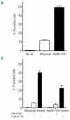
Rituximab-resistant cells are lysed by HuMab-7D8. (A) CD20 mAb-induced CDC of a CEM-CD20 clone with low CD20-expression (MFI of 325) by rituximab and HuMab-7D8. Cells were incubated with human serum only or with either rituximab or HuMab-7D8 and human serum. After 24 h the cells were washed and a sample was taken and analyzed for CDC. Results are expressed as % PI positive cells. The bars indicate the mean ± SD of triplicate measurements. (B) The cells that survived the one day initial rituximab- or Humab-7D8-mediated killing were cultured for an additional two weeks. Rituximab treated cells were subjected to a second exposure of CDC induction using either rituximab or HuMab-7D8. Conversely, the HuMab-7D8-treated cells from the first experiment were subjected to CDC induction using either rituximab or HuMab-7D8. Background lysis was assessed by adding normal human serum only.

Similar articles

Cited by

References

    1. Jemal A, Siegel R, Ward E, Murray T, Xu J, Thun MJ. Cancer statistics 2007. Cancer J Clin. 2007;57(1):43–66. - PubMed
    1. Marcus R, Hagenbeek A. The therapeutic use of rituximab in non-Hodgkin’s lymphoma. Eur J Haematol Suppl. 2007:5–14. - PubMed
    1. Groves FD, Linet MS, Travis LB, Devesa SS. Cancer surveillance series: non-Hodgkin’s lymphoma incidence by histologic subtype in the United States from 1978 through 1995. J Natl Cancer Inst. 2000;92(15):1240–51. - PubMed
    1. The Non-Hodgkin’s Lymphoma Classification Project: A clinical evaluation of the International Lymphoma Study Group classification of non-Hodgkin’s lymphoma. Blood. 1997;89(11):3909–18. - PubMed
    1. Fisher RI, Gaynor ER, Dahlberg S, Oken MM, Grogan TM, Mize EM, et al. Comparison of a standard regimen (CHOP) with three intensive chemotherapy regimens for advanced non-Hodgkin’s lymphoma. N Engl J Med. 1993;328(14):1002–6. - PubMed

Publication types

MeSH terms