. 2010 Nov 26;285(48):37704-15.
doi: 10.1074/jbc.M110.140517. Epub 2010 Sep 17.

Rapid fusion and syncytium formation of heterologous cells upon expression of the FGFRL1 receptor

Affiliations

Rapid fusion and syncytium formation of heterologous cells upon expression of the FGFRL1 receptor

Florian Steinberg et al. J Biol Chem. .

Abstract

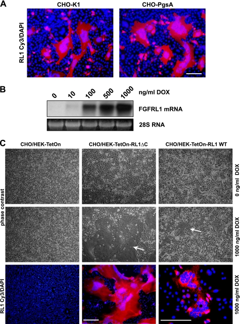
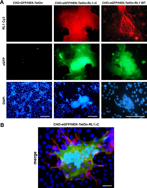
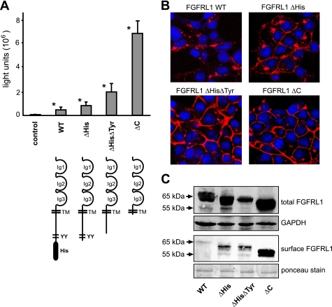
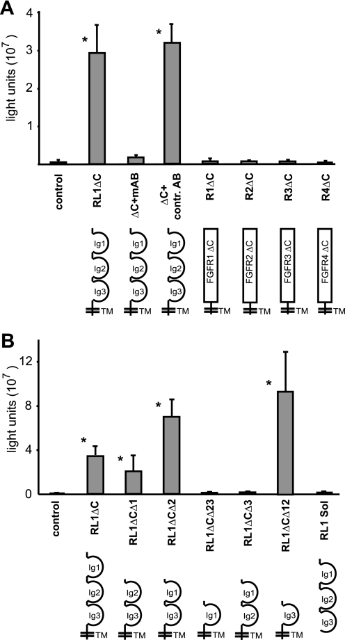
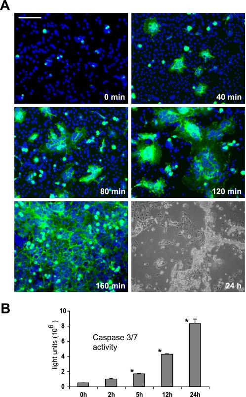
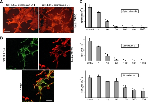
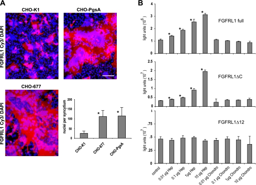
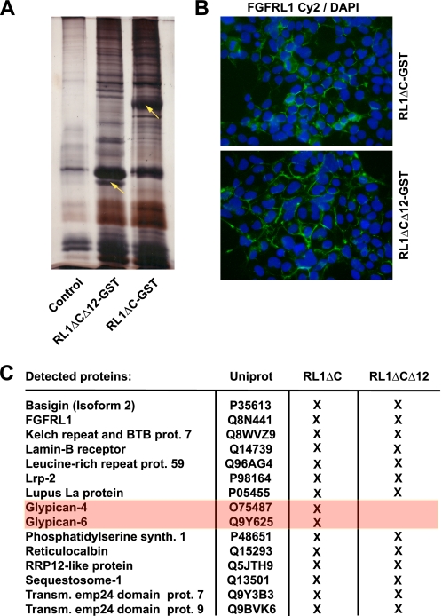
The fusion of mammalian cells into syncytia is a developmental process that is tightly restricted to a limited subset of cells. Besides gamete and placental trophoblast fusion, only macrophages and myogenic stem cells fuse into multinucleated syncytia. In contrast to viral cell fusion, which is mediated by fusogenic glycoproteins that actively merge membranes, mammalian cell fusion is poorly understood at the molecular level. A variety of mammalian transmembrane proteins, among them many of the immunoglobulin superfamily, have been implicated in cell-cell fusion, but none has been shown to actively fuse cells in vitro. Here we report that the FGFRL1 receptor, which is up-regulated during the differentiation of myoblasts into myotubes, fuses cultured cells into large, multinucleated syncytia. We used luciferase and GFP-based reporter assays to confirm cytoplasmic mixing and to identify the fusion inducing domain of FGFRL1. These assays revealed that Ig-like domain III and the transmembrane domain are both necessary and sufficient to rapidly fuse CHO cells into multinucleated syncytia comprising several hundred nuclei. Moreover, FGFRL1 also fused HEK293 and HeLa cells with untransfected CHO cells. Our data show that FGFRL1 is the first mammalian protein that is capable of inducing syncytium formation of heterologous cells in vitro.


Figures

FIGURE 1.
FIGURE 1.
FGFRL1 expression induces changes in cellular morphology that indicate cell-cell fusion and syncytium formation. A, CHO-K1 (left) and glycosaminoglycan deficient CHO-PgsA (right) cells were transfected with C-terminally truncated FGFRL1 (FGFRL1ΔC in pcDNA3.1). One day after transfection, cells were fixed and stained with a monoclonal antibody against FGFRL1 (Cy3, red) and DAPI. Note that most FGFRL1 positive CHO cells appear to be large, syncytial cell clusters with a continuous cytoplasm. Bar, 100 μm. B, Northern blot shows the induction of FGFRL1ΔC expression in a tetracycline inducible HEK-TetOn-RL1ΔC cell line with increasing concentrations of doxycycline. The cell line was derived from a single clone selected for tightly inducible expression. C, CHO-PgsA cells were seeded together with inducible, FGFRL1 wild type (right) and FGFRL1ΔC-expressing cell line (middle), or with regular HEK-TetOn cells (left) as a control. Upon addition of 1000 ng/ml doxycycline to the co-cultures, large “fusion plaques” (indicated by white arrows) appeared in the dishes with the FGFRL1-expressing HEK cell lines. No fusion was observed in the control dishes and in dishes without doxycycline. Antibody staining of FGFRL1 (Cy3, red) shows that the large cell aggregates uniformly expressed FGFRL1. Note that the image showing the fused FGFRL1 wild-type-expressing cells was taken at a higher magnification. Bar, 200 μm.
FIGURE 2.
FIGURE 2.
Fusion and cytoplasmic mixing of HEK and CHO cells upon expression of FGFRL1. A, CHO-PgsA cells were transfected with a tetracycline inducible eGFP expression construct, which is only active in the presence of the TetOn transactivator protein and doxycycline. The cells were trypsinized and seeded together with either HEK-TetOn-RL1ΔC, HEK-TetOn-RL1WT cells (express FGFRL1ΔC and FGFRL1 wild type, respectively, in the presence of doxycycline) or with regular HEK-TetOn cells. Doxycycline at 1000 ng/ml was added to all dishes. Note that both HEK-TetOn-RL1 cell lines fused with the CHO cells, leading to the diffusion of the TetOn transactivator from the HEK-TetON-RL1 cells into the syncytial cytoplasm. This activated the expression of eGFP in the large, syncytial cells. No fusion and no activation of eGFP expression was observed with the regular HEK-TetOn cells that served as controls. The image showing the fused FGFRL1 wild-type-expressing cells was taken at a higher magnification. Bar, 100 μm. B, higher powered magnification of a large, syncytial cell consisting of eGFP (green)-transfected CHO and FGFRL1ΔC (red)-expressing HEK-TetOn cells. Note that some FGFRL1-expressing HEK-TetOn cells (red) appear to be attracted by the syncytial cell. Bar, 20 μm.
FIGURE 3.
FIGURE 3.
Role of the cytoplasmic domain on the fusion process. A, CHO-K1 cells were transfected with a tetracycline-inducible luciferase expression construct (pTRE-Luc) and seeded together with HEK-TetOn cells that had been transfected with different C-terminally truncated FGFRL1 expression constructs (the corresponding proteins are schematically shown below the respective bars). The luciferase activity, measured 14 h postseeding, correlated well with the visible extent of cell-cell fusion induced by the mutated FGFRL1 constructs. The bars represent the average of three experiments. Successive truncation of cytoplasmic internalization motifs led to increased fusion, with strongest fusion observed for the completely truncated construct FGFRL1ΔC. Note that the wild-type, full-length protein also induces considerable fusion when compared with nontransfected control cells. Asterisks next to bars indicate statistically significant results (p < 0.05) compared with the control group. B, confocal images of immunofluorescent stainings of the C-terminally truncated FGFRL1 proteins. Note that the wild-type protein resided primarily in intracellular compartments, while the truncated forms accumulated in the plasma membrane. C, surface biotinylation of mutated FGFRL1 proteins. HEK293 cells stably expressing the indicated FGFRL1 proteins were subjected to surface biotinylation, followed by isolation with neutravidin agarose and detection of biotinylated FGFRL1 by Western blotting. The upper panel shows total FGFRL1 in the respective cells, with GAPDH as a loading control. The lower panel shows biotinylated FGFRL1 from the cell surface. Ponceau staining of a major membrane protein of ∼85 kDa served as a loading control for the neutravidin purification.
FIGURE 4.
FIGURE 4.
Fusogenic activity is specific to FGFRL1 and mediated by Ig-domain III. FGFRL1ΔC-transfected HEK-TetOn cells were co-cultured with pTRE-Luc-transfected CHO-K1 cells and allowed to fuse overnight, followed by measurement of luciferase activity. A, addition of a monoclonal antibody against the FGFRL1 ectodomain into the culture medium blocked fusion at 2 μg/ml. A control antibody against the intracellular domain of FGFRL1 (not present anymore in FGFRL1ΔC) had no effect. C-terminally truncated FGFR1-FGFR4 did not induce any cell-cell fusion in this cellular system. B, deletion of extracellular Ig-domains I and II leaves fusogenic activity intact. Deletion of Ig-domain III completely disrupts fusion. Note that both the RL1ΔCΔ2 and the RL1ΔCΔ12 proteins show increased activity when compared with the RL1ΔC construct. The soluble FGFRL1 ectodomain does not induce any cell-cell fusion. The asterisks indicate statistically significant results (p < 0.05).
FIGURE 5.
FIGURE 5.
FGFRL1ΔCΔ12 rapidly fuses HEK293 and CHO-PgsA cells and induces apoptotic cell death. A, kinetics of the FGFRL1-induced cell-cell fusion. HEK293 cells were transfected with FGFRL1ΔCΔ12-eGFP and seeded together with CHO-PgsA cells. The time of initial attachment was taken as the starting point. The figure displays snapshots taken every 40 min after the initial attachment of the cells. The first syncytial, multinucleated cells appeared after 40 min and rapidly expanded until, after 160 min, the entire field of vision was one continuous syncytium. 24 h after the onset of fusion, cells detached and appeared to be dying. Bar, 100 μm. B, activity of caspase 3/7 in the fusing HEK293 and CHO-PgsA cells. A luminometric caspase 3/7 activity kit was used to determine the activity of these effector caspases in the fusing cells. The asterisks indicate significant (p < 0.05) increases in caspase activity relative to unfused cells.
FIGURE 6.
FIGURE 6.
The actin skeleton is involved in FGFRL1-mediated cell-cell fusion. A, stable HEK-TetOn cell line with inducible expression of FGFRL1ΔC was cultured with (right) and without (left) doxycyline in the medium. After fixing of the cells, the actin cytoskeleton was stained with phalloidin-TRITC. FGFRL1 expression induces the formation of actin rich protrusions resembling filopodia. We also observed a redistribution of f-actin to the cell periphery. Bar, 8 μm. B, confocal image of antibody stained FGFRL1ΔC (green) and phalloidin-TRITC stained f-actin (red). The overlay shows that there is substantial overlap between FGFRL1ΔC and f-actin. Bar, 20 μm. C, luciferase-based cell-cell fusion assay was utilized to test the effect of the f-actin destabilizing compounds latrunculin-B and cytochalasin-D and of the microtubule disrupting agent nocodazole (all at 1–1000 ng/ml) on FGFRL1ΔC-induced cell-cell fusion. Either of the actin affecting drugs was strongly inhibitory on the fusogenic activity of FGFRL1ΔC and completely blocked fusion at higher concentrations. Nocodazole did not reduce the fusion at lower concentrations and moderately affected fusion at highly toxic concentrations. The bars represent the average luciferase activity measured in three wells. Asterisks indicate statistically significant (p < 0.05) differences relative to untreated control.
FIGURE 7.
FIGURE 7.
Cell surface heparan sulfates have an inhibitory effect on FGFRL1-mediated cell fusion. CHO-K1 cells, CHO-677 cells (lack heparan sulfates) and CHO-PgsA cells (lack all glycosaminoglycans) were seeded together with FGFRL1ΔC-transfected HEK-TetOn cells and left to fuse overnight. A, representative pictures of the resulting HEK/CHO syncytia are shown for each CHO cell line. Bar, 100 μm. The diagram gives the average number of nuclei per syncytium of the fusing CHO-K1, CHO-677, and CHO-PgsA cells with FGFRL1-transfected HEK cells. Both CHO-677 and CHO-PgsA cells displayed dramatically increased cell fusion efficiency when compared with the wild-type CHO-K1 cells, indicating that heparan sulfates have an inhibitory effect on FGFRL1-induced cell-cell fusion. B, luciferase (pTRE-Luc)-transfected CHO-K1 and FGFRL1 wild-type, FGFRL1ΔC-, and FGFRL1ΔCΔ12-transfected HEK-TetOn cells were co-cultured overnight in the absence and presence of exogenous heparin or chondroitin-4-sulfate in the culture medium, followed by luciferase measurement.
FIGURE 8.
FIGURE 8.
Mass spectrometric identification of proteins that co-purified with GST-tagged FGFRL1. A, GST-tagged FGFRL1 proteins were expressed in HEK293 cells (stable expression) and purified from Triton X-100 lysates with glutathione beads. Bound proteins were eluted from the beads with saturated urea. The silver-stained gel shows resolved proteins from a control pull-down (untransfected cells) versus proteins from FGFRL1ΔC-GST and FGFRL1ΔCΔ12-GST pull-downs from the transfected cells. The presence of additional silver-stained bands indicated that a number of proteins co-purified with the GST-tagged FGFRL1 proteins (yellow arrows indicate FGFRL1ΔC-GST and FGFRL1ΔCΔ12-GST). B, immunofluorescent staining of FGFRL1ΔC-GST and FGFRL1ΔCΔ12-GST proteins in the stably transfected HEK293 cells. Both constructs retained a plasma membrane localization. C, eluates were subjected to LC-MS shotgun sequencing to identify the proteins that were purified together with the FGFRL1 proteins. The table lists proteins that associated with FGFRL1ΔC-GST and FGFRL1ΔCΔ12-GST in HEK293 cells.

Similar articles

Cited by

References

    1. Chen E. H., Grote E., Mohler W., Vignery A. (2007) FEBS Lett. 581, 2181–2193 - PubMed
    1. Oren-Suissa M., Podbilewicz B. (2007) Trends Cell Biol. 17, 537–546 - PubMed
    1. Martens S., McMahon H. T. (2008) Nat. Rev. Mol. Cell Biol. 9, 543–556 - PubMed
    1. Sapir A., Choi J., Leikina E., Avinoam O., Valansi C., Chernomordik L. V., Newman A. P., Podbilewicz B. (2007) Dev. Cell 12, 683–698 - PMC - PubMed
    1. Podbilewicz B., Leikina E., Sapir A., Valansi C., Suissa M., Shemer G., Chernomordik L. V. (2006) Dev. Cell 11, 471–481 - PubMed

Publication types

Substances

LinkOut - more resources