. 2010 Nov 26;285(48):37762-72.
doi: 10.1074/jbc.M110.143099. Epub 2010 Sep 17.

A novel p53 phosphorylation site within the MDM2 ubiquitination signal: I. phosphorylation at SER269 in vivo is linked to inactivation of p53 function

Affiliations

A novel p53 phosphorylation site within the MDM2 ubiquitination signal: I. phosphorylation at SER269 in vivo is linked to inactivation of p53 function

Jennifer A Fraser et al. J Biol Chem. .

Abstract

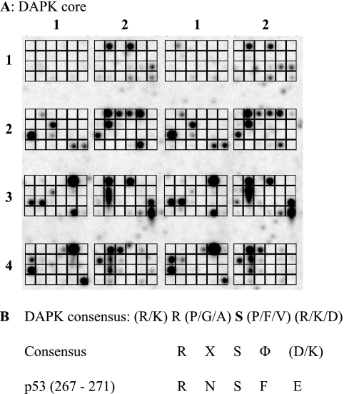
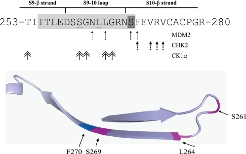
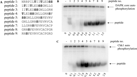
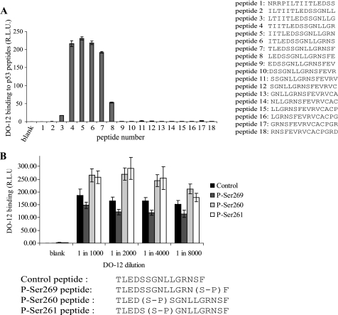
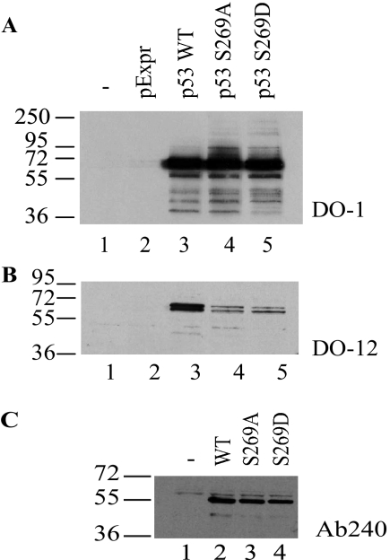
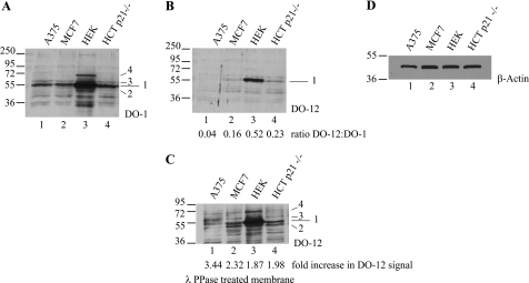
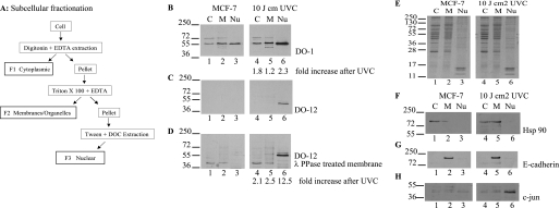
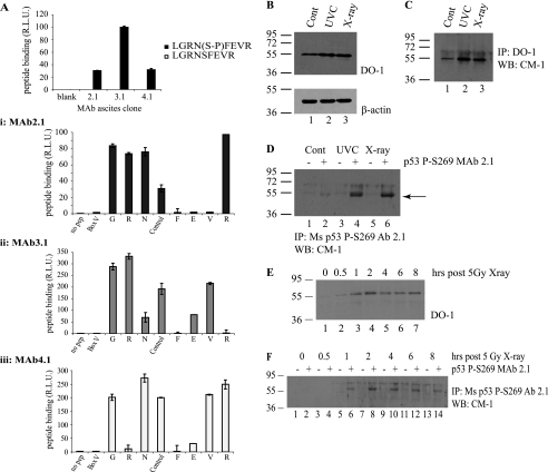
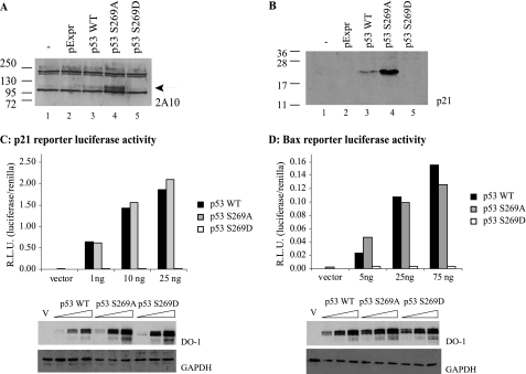
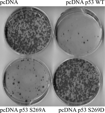
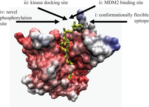
p53 is a thermodynamically unstable protein containing a conformationally flexible multiprotein docking site within the DNA-binding domain. A combinatorial peptide chip used to identify the novel kinase consensus site RXSΦ(K/D) led to the discovery of a homologous phosphorylation site in the S10 β-strand of p53 at Ser(269). Overlapping peptide libraries confirmed that Ser(269) was a phosphoacceptor site in vitro, and immunochemical approaches evaluated whether p53 is phosphorylated in vivo at Ser(269). Mutation or phosphorylation of p53 at Ser(269) attenuates binding of the p53-specific monoclonal antibody DO-12, identifying an assay for measuring Ser(269) phosphorylation of p53 in vivo. The mAb DO-12 epitope of p53 is masked via phosphorylation in a range of human tumor cells with WT p53 status, as defined by increased mAb DO-12 binding to endogenous p53 after phosphatase treatment. Phospho-Ser(269)-specific monoclonal antibodies were generated and used to demonstrate that p53 phosphorylation is induced at Ser(269) after irradiation with kinetics similar to those of p53 protein induction. Phosphomimetic mutation at Ser(269) inactivated the transcription activation function and clonogenic suppressor activity of p53. These data suggest that the dynamic equilibrium between native and unfolded states of WT p53 can be modulated by phosphorylation of the conformationally flexible multiprotein binding site in the p53 DNA-binding domain.


Figures

FIGURE 1.
FIGURE 1.
Pepchip kinase array screen identifies a novel phosphoacceptor site in the DNA-binding domain of p53. A, in vitro kinase consensus site definition using peptide arrays. A Pepchip array (total of 192 peptides) was incubated with 30 ng of DAPK core and [γ-33P]ATP for 3 h at 30 °C, and phosphorylated peptides were detected by storage phosphor screen. The array is arranged in a grid from 1 to 4 in the y axis and 1 and 2 in the x axis (in duplicate). B, consensus sites for protein kinases. Phosphorylated peptides from A (supplemental Table I) were analyzed as indicated in Table 1 to develop the consensus site summarized (RXSF(D/K)), which matches the Ser269 site in the p53 tumor suppressor protein.
FIGURE 2.
FIGURE 2.
The multiprotein docking sites within the p53 DNA-binding domain. Protein-protein contacts in the conformationally flexible ubiquitination signal from the DNA-binding domain of p53. The key residues involved in MDM2, Chk2, and CK1a binding (arrows) and the contact sites for the DO-12 monoclonal antibody (green) are highlighted. The bottom panel indicates the positioning of key residues in the region known to influence p53 stability.
FIGURE 3.
FIGURE 3.
The multiprotein docking peptide from the ubiquitin signal in the p53 DNA-binding domain contains an in vitro phosphoacceptor site. A, overlapping peptides corresponding to the conformationally flexible ubiquitination signal (with Ser260/261 phosphoacceptor sites in peptides 1–5 or Ser269 phosphoacceptor site in peptides 5–9) were incubated in kinase reactions containing [γ-32P]ATP and either DAPK core (B) or Chk1 (C) for 30 min at 30 °C. Reaction products were resolved via 20% SDS-PAGE and dried, and phosphopeptides were visualized by a PhosphorImager (highlighted by arrows). The intensity of peptide phosphorylation was quantified and normalized to kinase autophosphorylation and is expressed below each panel.
FIGURE 4.
FIGURE 4.
Attenuation of DO-12 epitope binding by Ser269 phosphorylation. A, DO-12 binding to the p53 DNA-binding domain. The region bound by the DO-12 monoclonal antibody was mapped by ELISA using overlapping peptides derived from the DNA-binding domain of p53 (A, right). B, serine 269 phosphorylation attenuates DO-12 binding. The effect of phosphorylation on the binding affinity of DO-12 to peptides (amino acids 255–270 of human p53) derived from the DNA-binding domain of p53 was defined by ELISA and is shown as a function of DO-12 antibody titration. The peptide sequences and modifications at serine 260, serine 261, or serine 269 are shown below. R.L.U., relative light units. Error bars, S.D.
FIGURE 5.
FIGURE 5.
Mutagenesis of full-length p53 at codon 269 attenuates DO-12 binding. H1299 cells were transfected with pExpr expression vectors encoding p53, p53S269A, or p53S269D mutants, and the proteins were examined for reactivity toward DO-1 (A; total p53), DO-12 (B; epitope 255–270), or PAb240 (C; epitope 209–214) by immunoblotting.
FIGURE 6.
FIGURE 6.
In vivo phosphorylation of endogenous p53 at serine 269; detection by phosphatase unmasking of antibody epitopes. A–C, phosphatase activity exposes the DO-12 epitope on endogenous p53. Lysates from A375, MCF-7, HEK-293, and HCT-116 p21−/− cells were resolved by electrophoresis, and immunoblots were probed with antibody for total p53 (A; DO-1), DO-12-reactive p53 (B), or DO-12-reactive p53 on blots pretreated with λ-phosphatase prior to incubation with DO-12 (C). The various p53-cross-reactive bands are indicated (1–4). Band intensity was quantified by Scion Image software, and the ratio of DO-12 to DO-1 signal and the degree of DO-12 unmasking are indicated below B and C, respectively. Equal sample loading was determined by immunoblotting with β-actin (D).
FIGURE 7.
FIGURE 7.
Serine 269-phosphorylated p53 has a selective subcellular localization. A, schematic representation of the fractionation methodology. B–D, effects of irradiation on p53 phosphorylation at the DO-12 epitope in MCF7 cells. MCF-7 cells were untreated or irradiated with UVC and grown for a further 6 h before fractionation into cytosolic (C), membrane/organelle (M), and nuclear fractions (Nu). Fractions were resolved by electrophoresis and immunoblotting with DO-1 (B) and DO-12 before (C) and after phosphatase treatment of the membrane (D) to define p53 localization. Band intensity was quantified by Scion Image software, and the -fold increase in antibody signal following UVC irradiation is indicated below B and D, respectively. Equivalent sample loading is shown by Coomassie staining of the various fractions (E), and fractionation of the distinct subcellular fractions was demonstrated by probing for known cytoplasmic (Hsp90), membrane (E-cadherin), and nuclear (c-Jun) marker proteins (F, G, and H, respectively).
FIGURE 8.
FIGURE 8.
In vivo phosphorylation of p53 at serine 269; detection using phosphospecific antibodies. A, phosphospecific antibody generation to serine 269. Monoclonal antibodies (mAb 2.1, 3.1, and 4.1) were generated toward phospho-Ser269 peptides, and peptide ELISA was used to demonstrate the phosphospecificity of the antibodies. A, i–iii, serine 269 mAb (i, mAb 2.1; ii, mAb 3.1; iii, mAb 4.1) epitopes were mapped via ELISA using an alanine scan mutation of GRNpSFEVR phosphopeptide. B–D, Ser269-phosphospecific mAb 2.1 can immunoprecipitate p53 from MCF7 lysates following DNA damage. MCF7 cells were exposed to 5 grays of x-ray or 10 J cm2 UV irradiation, and induction of p53 protein levels was analyzed 6 h later by immunoblotting with DO-1 (B, showing 10 μg of cellular lysate probed with DO-1 (top) and β-actin (bottom) as a loading control). Total p53 protein and Ser269-phosphorylated p53 were isolated from these lysates by immunoprecipitation with DO-1 (C) or mAb 2.1 (D), respectively, and an increase in Ser269 phosphorylation can be detected (D, lanes 4 and 6). Protein G beads were used as a control for both DO-1 and mAb 2.1 immunoprecipitation and are shown in D in lanes marked with minus signs. This increase in phosphorylation with phosphospecific antibody after irradiation is consistent with the pool of DO-12 non-reactive p53 protein in the nucleus of cells after UV radiation (Fig. 7, C versus D). E–F, time scale of p53 Ser269 phosphorylation following DNA damage. MCF-7 cells were irradiated with 5 grays of x-ray radiation and harvested at the indicated time points. Increases in total p53 (E) or phospho-Ser269 p53 (F) protein levels were determined by immunoblotting cellular lysates with DO-1 (E) or immunoprecipitating phospho-Ser269 p53 from cellular lysates using protein G beads without (−) or with mAb 2.1 (+), followed by immunoblotting with polyclonal CM-1 (F). R.L.U., relative light units; IP, immunoprecipitation; IB, immunoblot.
FIGURE 9.
FIGURE 9.
The effects of codon 269 mutation on p53 activity in cells; phosphomimetic substitution of serine 269 inactivates p53. The effect of serine 269 mutation on expression of the p53 downstream target genes, Mdm2 and p21, was examined by immunoblotting 20 μg of lysates derived from H1299 cells transfected with pcDNA vector control, wild type p53, p53S269A, or p53S269D mutants using the anti-MDM2 monoclonal antibody 2A10 (A) or the p21 monoclonal antibody Ab-1 (B). C and D, p53 function from reporters containing p53-responsive binding sites. H1299 cells were transfected with the p21 or Bax reporter plasmids alone or with (1–75 ng of) pExpr p53, S269A, or S269D expression vectors. Total DNA was normalized with pExpr vector control plasmid DNA. Twenty-four hours later, cells were lysed, and luciferase activity from p21 (C) or Bax (D) reporters was determined using dual luciferase reporter assays. Data were normalized by expressing luciferase activity (luciferase/Renilla) in relative light units (R.L.U.). The lower panels depict p53 protein levels after transfection by immunoblotting. GAPDH immunoblotting was used as a loading control.
FIGURE 10.
FIGURE 10.
Phosphomimetic mutation of Ser269 inhibits p53 growth-suppressive activity. H1299 cells were transfected with 1 μg of pcDNA plasmid containing wild type, S269A, or S269D. Twenty-four hours later, cells were trypsinized and diluted and then plated in medium containing 0.5 mg/ml Geneticin and grown for a further 2 weeks. Colonies were fixed and stained with Giemsa dye.
FIGURE 11.
FIGURE 11.
Features of the regulatory motif in the p53 DNA-binding domain. The multiprotein binding site in the p53 core DNA-binding domain (highlighted in yellow) has a conformationally flexible motif (24) (i), MDM2-docking site or ubiquitination signal (22, 40) (ii), kinase docking site (25, 26, 29) (iii), and novel phosphorylation site at Ser269 (this study) (iv).

Similar articles

Cited by

References

    1. Horn H. F., Vousden K. H. (2004) Nature 427, 110–111 - PubMed
    1. Brooks C. L., Gu W. (2006) Mol. Cell. 21, 307–315 - PMC - PubMed
    1. Dornan D., Shimizu H., Burch L., Smith A. J., Hupp T. R. (2003) Mol. Cell. Biol. 23, 8846–8861 - PMC - PubMed
    1. Dornan D., Hupp T. R. (2001) EMBO Rep. 2, 139–144 - PMC - PubMed
    1. Lambert P. F., Kashanchi F., Radonovich M. F., Shiekhattar R., Brady J. N. (1998) J. Biol. Chem. 273, 33048–33053 - PubMed

Publication types