. 2010 Nov 19;285(47):36532-41.
doi: 10.1074/jbc.M110.169029. Epub 2010 Sep 17.

The archaeal topoisomerase reverse gyrase is a helix-destabilizing protein that unwinds four-way DNA junctions

Affiliations

The archaeal topoisomerase reverse gyrase is a helix-destabilizing protein that unwinds four-way DNA junctions

Anna Valenti et al. J Biol Chem. .

Abstract

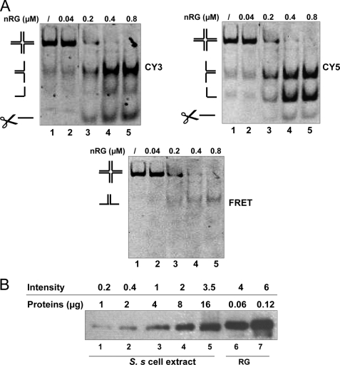
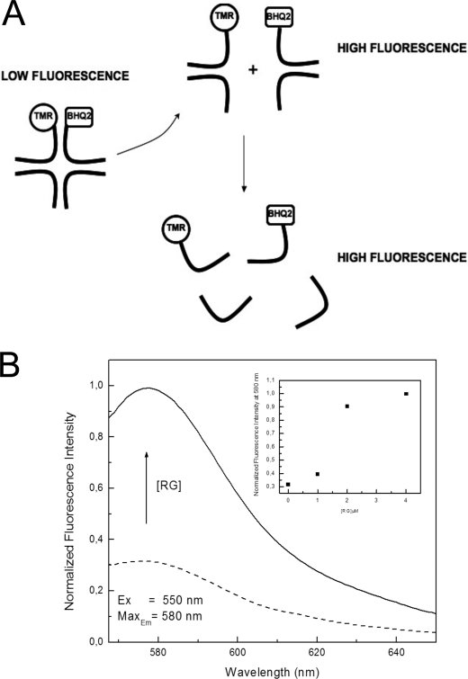
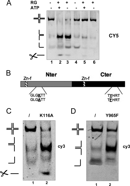
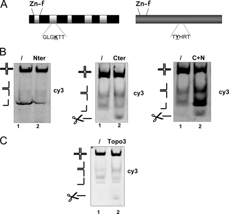
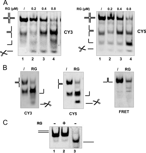
Four-way junctions are non-B DNA structures that originate as intermediates of recombination and repair (Holliday junctions) or from the intrastrand annealing of palindromic sequences (cruciforms). These structures have important functional roles but may also severely interfere with DNA replication and other genetic processes; therefore, they are targeted by regulatory and architectural proteins, and dedicated pathways exist for their removal. Although it is well known that resolution of Holliday junctions occurs either by recombinases or by specialized helicases, less is known on the mechanisms dealing with secondary structures in nucleic acids. Reverse gyrase is a DNA topoisomerase, specific to microorganisms living at high temperatures, which comprises a type IA topoisomerase fused to an SF2 helicase-like module and catalyzes ATP hydrolysis-dependent DNA positive supercoiling. Reverse gyrase is likely involved in regulation of DNA structure and stability and might also participate in the cell response to DNA damage. By applying FRET technology to multiplex fluorophore gel imaging, we show here that reverse gyrase induces unwinding of synthetic four-way junctions as well as forked DNA substrates, following a mechanism independent of both the ATPase and the strand-cutting activity of the enzyme. The reaction requires high temperature and saturating protein concentrations. Our results suggest that reverse gyrase works like an ATP-independent helix-destabilizing protein specific for branched DNA structures. The results are discussed in light of reverse gyrase function and their general relevance for protein-mediated unwinding of complex DNA structures.


Figures

FIGURE 1.
FIGURE 1.
Gel-FRET assay for HJ processing by RG. A, a model of the assay. HJ was assembled by annealing the A1-A4 oligonucleotides shown in Table 1. The 5′-end of strand A2 and the 3′-end of strand A1 were labeled with Cy5 and Cy3 fluorophores, respectively. The graphic shows HJ and predicted junction unfolding products. B, Cy5/Cy3-labeled HJ (20 nm) was incubated at 55 °C for 30 min without (lane 1) or with increasing concentrations of RG as indicated (lanes 2–5). Samples were run, and the same gel was scanned under different excitation/emission conditions as described under “Experimental Procedures.” The panels show the images obtained for Cy3, Cy5, and FRET and the merge of all three images, respectively. Reaction substrate and products (forks, ss, and cleavage products, which are indicated by scissors) are indicated. The complicated band pattern seen in the merge panel is due to different rates of migration of the two fluorophore-labeled oligonucleotides with each other and with respect to the unlabeled ones. The gel is representative of 20 independent experiments obtained with three different protein preparations.
FIGURE 2.
FIGURE 2.
HJ processing by nRG. A, gel-FRET assay for HJ processing by nRG. HJ (20 nm) was incubated for 30 min at 55 °C without (lane 1) or with increasing concentrations of nRG as indicated (lanes 2–5). Gel electrophoresis and analysis were as described in the legend for Fig. 1. The panels show the images obtained for Cy3, Cy5, and FRET, respectively. The gel is representative of three independent experiments. B, quantification of reverse gyrase in cell extract. Soluble S. solfataricus cell extracts (S. s cell extract, lanes 1–5) and purified RG (lanes 6 and 7) were probed with the anti-TopR1 polyclonal antibody (19) used at 1:10,000 dilution. This antibody recognizes both recombinant TopR1 and recombinant TopR2 (see Footnote 3), the two in S. solfataricus reverse gyrase isoforms. The intensity of the bands, determined using the VersaDoc instrument (Bio-Rad) and expressed as arbitrary units, is reported above the amount of protein loaded in each lane. The intensity of the reverse gyrase band in lane 5 (16 μg of cell extract) corresponds roughly to that obtained with 0.06 μg of purified protein (lane 6); thus, reverse gyrase accounts for about 0.37% of total soluble protein.
FIGURE 3.
FIGURE 3.
Fluorescence measurements of HJ. A, Q-HJ was assembled by annealing the A3-A4-A8-A9 oligonucleotides shown in Table 1. The 3′-end of strand A8 and 5′-end of strand A9 were labeled with the TAMRA fluorophore (TMR) and the BHQ2 quencher, respectively. The graphic shows the model of the assay. B, fluorescence emission spectra were recorded as described under “Experimental Procedures.” In each reaction, 0.1 μm Q-HJ was used. Plots of the change in fluorescence intensity on the y axis (arbitrary units), in the absence (dashed lines) or presence of 4.0 μm RG (black lines), are shown. The inset shows the effect of increasing RG concentrations on the fluorescence intensity at 580 nm (y axis); the plateau was reached between 2.0 and 4.0 μm. Ex, excitation; Max, maximum.
FIGURE 4.
FIGURE 4.
RG catalytic activities are dispensable for HJ processing. A, HJ (20 nm) was incubated at 55 (lanes 1–3) or 37 °C (lanes 4–6) for 30 min without (lanes 1 and 4) or with (lanes 2, 3, 5, and 6) RG at 0.8 μm; in lanes 2 and 5, 5 mm ATP was added. Only the scan from CY5 emission is shown. The gel is representative of three independent experiments. B, schematic diagram of RG showing the two domains, the point mutations in the ATP-binding site of the N-terminal domain (Nter) and in the catalytic tyrosine of the C-terminal domain (Cter), and the position of the two putative zinc finger motifs (Zn-f) for DNA binding (19). C and D, gel-FRET assays. HJ (20 nm) was incubated under standard conditions without (lane 1) or with (lane 2) reverse gyrase mutants, as indicated. All proteins were used at 0.8 μm; only the scans from CY3 emission are shown. The gels are representative of four independent experiments performed with two independent mutant proteins preparations.
FIGURE 5.
FIGURE 5.
Isolated RG domains are inactive in HJ processing. A, schematic diagram showing the RG N-terminal and C-terminal domains. The two putative zinc finger motifs (Zn-f) for DNA binding are indicated. B, gel-FRET assays. HJ (20 nm) was incubated under standard conditions without (lane 1) or with (lane 2) reverse gyrase mutants, as indicated. All proteins were used at 0.8 μm; only the scans from Cy3 emission are shown. The gels are representative of three independent experiments. Nter, N-terminal domain; Cter, C-terminal domain; C+N, C- and N-terminal domains. C, HJ (20 nm) was incubated under standard conditions without (lane 1) or with (lane 2) E. coli Topo3 (0.8 μm). The gels are representative of three independent experiments performed.
FIGURE 6.
FIGURE 6.
Substrates of ATP-independent unwinding. A, processing of immobile HJ. IM-HJ (20 nm) was produced by annealing the A1, A2, A5, and A6 oligonucleotides shown in Table 1 and was incubated at 55 °C for 30 min without (lane 1) or with increasing concentrations of RG, as indicated (lanes 2–4). The panels show the images obtained for Cy3 and Cy5, respectively. The gel is representative of two independent experiments. B, ATP-independent unwinding of a fork substrate. A gel-FRET assay with fork substrate (oligonucleotides A1+A2 in supplemental Table S1, 20 nm) and RG (0.8 μm) is shown. Incubation was for 30 min at 55 °C. The panels show the images obtained for Cy3, Cy5, and FRET, respectively. The gel is representative of four independent experiments. C, RG does not unwind a ds substrate. A gel-FRET assay was performed with the 40-bp ds substrate (oligonucleotides A2+A7 in Table 1, 20 nm) incubated without (lane 1) or with RG (0.8 μm; lane 2) at 55 °C for 30 min. Lane 3, denatured ds. Only the scan from Cy5 emission is shown. The gel is representative of two independent experiments.
FIGURE 7.
FIGURE 7.
Binding of RG to HJ distorts its structure. A, RG binding to HJ was analyzed by EMSA. Increasing concentrations of RG (60, 120, 240, 480 nm) were incubated at 37 °C with the following 32P-labeled substrates (20 nm): HJ, 40-bp ds (oligonucleotide A2+A7 in supplemental Table S1), fork (oligonucleotides A1+A2 in supplemental Table S1), and 80-bp ds (Valenti et al. 21). The graph shows the quantification of results expressed as the percentage of shifted DNA versus the P/DNA ratio. For each DNA ligand, the fraction of shifted DNA versus the amount of protein used is plotted. Binding assays were performed in triplicate, and the results were averaged. Values are the mean ± S.E. of three independent experiments. B, HJ probing by potassium permanganate. 32P-labeled-HJ (20 nm) was incubated with RG at 0.6 μm (lanes 1 and 3) or without protein (lanes 2 and 4) at 37 °C for 30 min; after incubation, samples were reacted with KMnO4 followed by piperidine cleavage (lane 1 and 2) or directly incubated with piperidine (lane 3); lanes 5 and 6 show molecular weight markers of 22 and 18 nucleotides, respectively. Controls were mock-treated exactly as samples. The gel is representative of three independent experiments. C, the two extreme conformations of HJ with its 4-base homologous core (underlined). Arrows indicate the thymines specifically cleaved by piperidine after KMnO4 treatment in the presence of RG.

References

    1. Sharples G. J. (2001) Mol. Microbiol. 39, 823–834 - PubMed
    1. Lilley D. M. (2010) Biochem. Soc. Trans. 38, 399–403 - PubMed
    1. Briggs G. S., Mahdi A. A., Weller G. R., Wen Q., Lloyd R. G. (2004) Philos. Trans. R. Soc. Lond. B Biol. Sci. 359, 49–59 - PMC - PubMed
    1. Whitby M. C. (2010) DNA Repair 9, 224–236 - PubMed
    1. Vindigni A., Hickson I. D. (2009) HFSP J. 3, 153–164 - PMC - PubMed

Publication types

MeSH terms

LinkOut - more resources