. 2010 Nov;192(22):6025-38.
doi: 10.1128/JB.00654-10. Epub 2010 Sep 17.

Calcium and iron regulate swarming and type III secretion in Vibrio parahaemolyticus

Affiliations

Calcium and iron regulate swarming and type III secretion in Vibrio parahaemolyticus

Cindy J Gode-Potratz et al. J Bacteriol. 2010 Nov.

Abstract

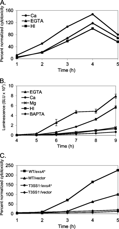
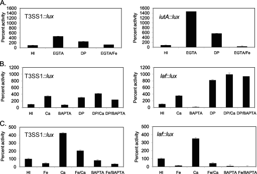
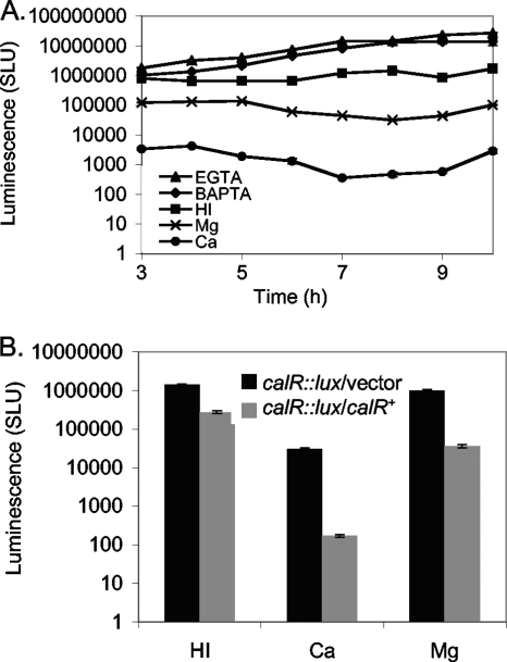
Here, we probe the response to calcium during growth on a surface and show that calcium influences the transcriptome and stimulates motility and virulence of Vibrio parahaemolyticus. Swarming (but not swimming) gene expression and motility were enhanced by calcium. Calcium also elevated transcription of one of the organism's two type III secretion systems (T3SS1 but not T3SS2) and heightened cytotoxicity toward host cells in coculture. Calcium stimulation of T3SS gene expression has not been reported before, although low calcium is an inducing signal for the T3SS of many organisms. EGTA was also found to increase T3SS1 gene expression and virulence; however, this was demonstrated to be the consequence of iron rather than calcium chelation. Ectopic expression of exsA, encoding the T3SS1 AraC-type regulator, was used to define the extent of the T3SS1 regulon and verify its coincident induction by calcium and EGTA. To begin to understand the regulatory mechanisms modulating the calcium response, a calcium-repressed, LysR-type transcription factor named CalR was identified and shown to repress swarming and T3SS1 gene expression. Swarming and T3SS1 gene expression were also demonstrated to be linked by LafK, a σ(54)-dependent regulator of swarming, and additionally connected by a negative-feedback loop on the swarming regulon propagated by ExsA. Thus, calcium and iron, two ions pertinent for a marine organism and pathogen, play a signaling role with global consequences on the regulation of gene sets that are relevant for surface colonization and infection.


Figures

FIG. 1.
FIG. 1.
Calcium regulates swarming motility and lateral flagellar gene expression. (A) Calcium affects rate of swarming. Strain LM5674 was spotted in the center of HI plates or HI plates supplemented with 4 mM CaCl2. Rates of radial expansion were calculated by periodically measuring the diameter of the swarming colony. Error bars represent standard deviations for at least 4 replica plates at each time point from one representative experiment. The slopes were calculated over the linear portion for each condition, from 3 to 7 h. The rate of expansion was 4.5 mm/h (R2 = 0.99) on HI and 6.3 mm/h (R2 = 0.99) on HI Ca. (B) Calcium affects laf::lux expression. Strain LM5738, with a lux fusion in a lateral flagellar gene (laf::lux), was inoculated on HI plates or HI plates supplemented with 4 mM CaCl2, 4 mM MgCl2, or 4 mM BAPTA. Plates were harvested periodically by suspending the cells to measure luminescence. Luminescence measured for each growth condition with supplement was calculated to be significantly different from that for growth in HI without supplement from 6 to 9 h (P < 0.001). (C) Ion specificity and laf::lux expression. Light production by LM5738 harvested from plates with the indicated CaCl2 or MgCl2 concentration was determined at 8 h. The statistical significance of the differences with and without supplement was calculated at P values of <0.0001 for 4 and 10 mM Ca and <0.001, <0.002, and <0.03 for 4, 10, and 40 mM Mg, respectively. The differences elicited by 40 and 4 mM Mg compared to 10 mM Mg were not considered statistically significant (P > 0.07). Luminescence is expressed as SLU, which are total light units per min normalized to OD600. Error bars represent standard deviations for triplicate light measurements from a representative experiment. Experiments were repeated at least 3 times.
FIG. 2.
FIG. 2.
Microarray expression profiles of selected genes. LM5674 was grown on HI plates or HI plates with 4 mM CaCl2 or 4 mM EGTA. Expression levels are log2 values of the normalized expression value for each gene under each condition. Locus tag designations are given, with the predicted products in parentheses. Error bars represent the standard errors from two replicates for each condition.
FIG. 3.
FIG. 3.
Calcium, EGTA, and ExsA induce T3SS1 cytotoxicity and gene expression. (A) Ca and EGTA affect cytotoxicity. Strain LM5191 was grown and harvested from HI plates or plates with 4 mM CaCl2 or 4 mM EGTA, as per the microarray growth conditions, and used to infect Chinese hamster ovary (CHO) cells at an MOI of 15. Cytotoxicity was assayed by measuring release of host cell lactate dehydrogenase (LDH) at 1-h intervals postinfection and is expressed as percent lysis normalized to the maximum lysis for LM5191 grown on HI. Values for the Ca and EGTA conditions were significantly different from those for HI at 3 to 5 h (P < 0.0006). (B) T3SS1::lux expression. Strain LM9649 carrying the T3SS1 reporter plasmid (with T3SS1::lux) was grown on HI kanamycin plates or HI kanamycin plates with 4 mM CaCl2, 4 mM MgCl2, 4 mM EGTA, or 4 mM BAPTA. Plates were harvested periodically by suspending the cells to measure luminescence. Luminescence is expressed as SLU from one representative experiment. Error bars represent the standard deviations for triplicate measurements at each time point. Values for each condition with supplement (Ca, EGTA, and BAPTA) were significantly different from those for HI at 6 to 9 h (P < 0.003); the Mg condition was not significantly different from HI. (C) ExsA affects cytotoxicity. The effect of exsA expression on cytotoxicity was measured by using strains grown and harvested as per the microarray experiment to infect CHO cells at an MOI of 15. The cytotoxicity of wild-type LM5674 carrying the exsA expression plasmid (pLM3650) or the T3SS1 mutant strain LM7035 (ΔVP1672::Camr) carrying the vector (pLM1877) or the exsA expression plasmid was significantly different from that of the wild-type strain carrying the vector at 4 and 5 h (P < 0.001). Cytotoxicity is expressed as percent lysis normalized to the maximum lysis for LM5674 with the vector. For the cytotoxicity experiments with results shown in panels A and C, error bars indicate standard errors of the means from 5 individual samples at each time point for a representative experiment. All experiments were repeated at least three times.
FIG. 4.
FIG. 4.
ExsA inhibits lateral flagellar gene expression and swarming motility. (A) ExsA affects laf::lux expression. The lateral flagellar gene reporter fusion strain LM5738 (laf::lux) carrying the vector or the IPTG-inducible exsA clone was grown on HI gentamicin plates with 1 mM IPTG and harvested periodically to measure luminescence, which is reported as SLU. Error bars represent standard deviations for triplicate light measurements from each sample. Light values for these strains were significantly different from each other at 5 to 7 h (P < 0.001). A representative experiment of three is shown. (B) ExsA affects swarming. Wild-type (WT) LM5674 or T3SS1 mutant strain LM7035 (ΔVP1672::Camr) carrying the vector or an IPTG-inducible exsA expression construct was spotted on HI gentamicin swarm plates with IPTG and incubated overnight.
FIG. 5.
FIG. 5.
Iron and calcium affect lateral flagellar and T3SS1 gene expression. (A) EGTA limits iron availability. (B) Effects of limiting iron in the presence or absence of calcium. (C) Effects of excess iron in the presence or absence of calcium. Reporter strains were grown on HI plates or plates with 4 mM EGTA, 50 μM FeCl3, 100 μM 2,2′-dipyridyl (DP), 4 mM CaCl2, or 4 mM BAPTA. Plates were harvested periodically, and results from one time point (9 h) are shown; however, trends were consistent throughout the time course. Reporter gene activity is expressed as the percent luminescence (calculated as SLU) produced under a given growth condition normalized to that produced under the HI condition. Error bars represent the standard deviations from triplicate measurements. The reporters were in strains LM9649 (T3SS1::lux), LM5738 (laf::lux), and LM6151 (ferric aerobactin receptor, iutA::lux). The experiments were repeated three times. All conditions with supplements elicited reporter expression significantly different from that with HI alone (P < 0.03), except for the EGTA/Fe and BAPTA effects compared to the HI effects for T3SS::lux, which were not considered different. The differences in expression for each reporter between EGTA and EGTA with Fe, Ca and Ca with Fe, and BAPTA and BAPTA with Fe were significant (P < 0.001).
FIG. 6.
FIG. 6.
The LysR-type transcription factor CalR is calcium regulated. (A) calR::lux expression. Strain LM7120 (calR::lux) was inoculated on HI plates and HI plates with 4 mM CaCl2, 4 mM MgCl2, 4 mM EGTA, or 4 mM BAPTA. Cells were harvested periodically to measure luminescence, reported as SLU. Error bars represent standard deviations from triplicate samples for one representative experiment of three. calR::lux expression levels were significantly different between HI and all other conditions from 6 to 9 h (P < 0.001). (B) CalR autoregulation. Luminescence is shown for LM7915 (calR::lux calR+) and LM7916 (calR::lux vector) grown in HI broth (with gentamicin and 50 μM IPTG) alone or supplemented with 4 mM CaCl2 or 4 mM MgCl2. calR was supplied in trans by using the IPTG-inducible plasmid pLM2795. Error bars represent standard deviations from triplicate samples for one representative experiment of three. For each growth condition, calR::lux expression levels were significantly different between strains carrying the calR expression plasmid and strains carrying the vector (P < 0.001).
FIG. 7.
FIG. 7.
CalR inhibits cytotoxicity, T3SS1 gene expression, swarming motility, and laf gene expression. (A) calR mutant cytotoxicity. Strain LM5191 and the congenic calR mutant LM5197 were grown on HI plates with or without 4 mM CaCl2 and used to infect CHO cells at an MOI of 15. Cytotoxicity is reported as percent lysis normalized to the maximum lysis for the wild type grown on HI. Error bars represent standard errors of the means from 5 replicates for one representative experiment of three. At 2 to 4 h postinfection, levels of LDH release were significantly different between the wild type grown on HI and all other conditions (P < 0.008). (B) calR mutant T3SS1::lux expression. Strains LM9649 and LM9650 (constructed from LM5191 and LM5197, respectively), each carrying the T3SS1::lux reporter on a plasmid, were grown on HI plates with or without 4 mM CaCl2. Luminescence was measured periodically. Error bars represent standard deviations from 3 light measurements for one representative experiment of three. At 6 to 9 h, T3SS1::lux expression levels were significantly different between the wild-type strain grown in calcium and grown in HI, between the calR strain grown in calcium and grown in HI, and between the calR strain and the wild-type strain grown in HI or HI Ca (P < 0.001). (C) calR mutant swarming rates. Strains used as described for panel A were spotted in the center of HI swarm plates with or without 4 mM CaCl2. Colony expansion was measured periodically. Error bars represent standard deviations from at least 4 replicates at each point for one representative experiment of three. Rates of radial expansion on HI Ca were 7.1 mm/h (R2 = 0.99) for LM5197 (calR) and 6.9 mm/h (R2 = 0.99) for LM5191 (WT). Rates on HI were 5.7 mm/h (R2 = 0.99) for LM5197 and 3.9 mm/h (R2 = 0.99) for LM5191. (D) calR mutant laf::lacZ expression. Strain LM5949 (laf::lacZ) and strain LM7120 (laf::lacZ calR::Tn5 lux) were grown on HI plates with or without 4 mM CaCl2. β-Galactosidase activity was measured periodically and is expressed in Miller units. From 6 to 9 h, LacZ activity for the wild type grown on Ca and the calR mutant grown on HI or HI Ca was significantly different from that for the wild type grown on HI (P < 0.001). Error bars represent standard deviations from triplicate assays for one representative experiment of three.
FIG. 8.
FIG. 8.
Calcium-enhanced cytotoxicity and T3SS1 gene expression require LafK. (A) lafK mutant cytotoxicity. Strain LM5674 and the congenic lafK mutant LM7789 were grown on HI plates or HI plates with 4 mM CaCl2 or 10 mM CaCl2. Cells were suspended in HI broth from these plates and used to infect CHO cells at an MOI of 15. Cytotoxicity, measured in the LDH release assay, is reported as percent lysis normalized to the maximum lysis for the wild type grown on HI. Error bars represent standard errors from 5 replicates for one representative experiment of three. From 3 to 5 h postinfection, the lafK mutant cytotoxicity was statistically different from that for the wild type grown under each condition (P < 0.01). (B) T3SS1::lux in the lafK mutant. T3SS1 gene expression was measured in strain LM5191 and the congenic lafK mutant strain LM9596, carrying a plasmid with a T3SS1::lux reporter. Strains were grown on HI plates with tetracycline (to maintain the reporter) or HI plates with 4 mM CaCl2 or 10 mM CaCl2. Cells were harvested periodically for luminescence measurements, expressed as SLU. Error bars represent standard deviations from triplicate assays at each time point for one representative experiment of three. From 7 to 9 h, expression in the lafK mutant was statistically different from expression in the wild type under each condition (P < 0.001).
FIG. 9.
FIG. 9.
Calcium regulates swarming and type III secretion. The availability of calcium affects global gene expression positively and negatively in V. parahaemolyticus. Two large gene sets displaying calcium-induced gene expression encode the swarming and T3SS1 regulons. This regulation is mediated in part by the calcium-repressed, LysR-type transcription factor CalR. Calcium represses calR transcription in CalR-dependent and -independent manners. calR mutants show enhanced swarming motility, laf gene expression, cytotoxicity, and T3SS1 gene expression. As a consequence, calR is repressed in high calcium, and swarming and virulence increase. The swarming regulator LafK is required to mediate a major portion of the T3SS1 response to calcium. In addition, evidence suggests a capacity for feedback regulation of swarming upon upregulation of exsA expression. Thus, during growth on a surface, calcium seems to act as a colonization signal influencing the capacity to swarm and be pathogenic; however, regulatory mechanisms also exist to link and modulate the expression of swarming and T3SS1 genes. Although not depicted, iron availability has been identified as an additional signal operating independently to influence expression of these two regulons; iron-limiting conditions induce swarming and T3SS1.

Similar articles

Cited by

References

    1. Belas, R., M. Simon, and M. Silverman. 1986. Regulation of lateral flagella gene transcription in Vibrio parahaemolyticus. J. Bacteriol. 167:210-218. - PMC - PubMed
    1. Bilecen, K., and F. H. Yildiz. 2009. Identification of a calcium-controlled negative regulatory system affecting Vibrio cholerae biofilm formation. Environ. Microbiol. 11:2015-2029. - PMC - PubMed
    1. Boles, B. R., and L. L. McCarter. 2000. Insertional inactivation of genes encoding components of the sodium-type flagellar motor and switch of Vibrio parahaemolyticus. J. Bacteriol. 182:1035-1045. - PMC - PubMed
    1. Brutinel, E. D., and T. L. Yahr. 2008. Control of gene expression by type III secretory activity. Curr. Opin. Microbiol. 11:128-133. - PMC - PubMed
    1. Butler, A. 2005. Marine microbial iron mobilization: new marine siderophores, abstr. U893. Abstr. Papers Am. Chem. Soc., vol. 229.

Publication types

MeSH terms

Associated data