. 2011 Feb 15;183(4):500-10.
doi: 10.1164/rccm.201005-0792OC. Epub 2010 Sep 17.

Dysregulation of p38 and MKP-1 in response to NOD1/TLR4 stimulation in sarcoid bronchoalveolar cells

Affiliations

Dysregulation of p38 and MKP-1 in response to NOD1/TLR4 stimulation in sarcoid bronchoalveolar cells

Ruchi Rastogi et al. Am J Respir Crit Care Med. .

Abstract

Rationale: Sarcoidosis is a systemic inflammatory disorder characterized by distinct up-regulation of Th1 cytokines, such as tumor necrosis factor (TNF)-α and IL-12. The mechanism underlying this up-regulation remains unclear. Recognition of microbial moieties through Toll-like or Nod-like receptors evokes sequential activation of mitogen-activated protein kinases (MAPKs), which plays a role in Th1-immune response.

Objectives: To test the hypothesis that dysregulation in MAPK signaling in response to microbial stimulation is important in mediating Th1 response in sarcoidosis.

Methods: Ex vivo cultured bronchoalveolar lavage (BAL) cells isolated from patients with sarcoidosis and control subjects were stimulated with low-dose Toll-like receptor 4 (TLR4) and nucleotide-binding oligomerization domain 1 (NOD1) ligands as a model of microbial stimulation, and MAPK signaling and inflammatory response were analyzed.

Measurements and main results: BAL cells from patients with sarcoidosis exhibited higher basal p38 activity, greater p38 phosphorylation, and more robust production of TNF-α and IL-12/IL-23p40 on stimulation with NOD1 and TLR4 agonists than cells isolated from control subjects. In contrast, control BAL cells had greater basal extracellular signal-regulated kinase (ERK) activity and NOD1 and TLR4 agonists preferentially activated the ERK pathway. Inhibition of p38, but not ERK, attenuated production of both IL12/IL23p40 and TNF-α. Interestingly, stimulation of cells from patients with sarcoidosis with either NOD1 or TLR4 ligand failed to induce MAPK phosphatase 1 (MKP-1). Adenovirus-mediated overexpression of MKP-1 attenuated p38 activation and decreased the production of IL12/IL23p40 and TNF-α in sarcoid BAL cells.

Conclusions: Our results suggest that enhanced p38 signaling in response to microbial products is caused by abnormal regulation of MKP-1 and contributes to heightened inflammation in sarcoidosis.


Figures

Figure 1.
Figure 1.
Baseline cytokine profiles of cultured bronchoalveolar lavage (BAL) cells from patients and control subjects. Cells obtained during BAL from patients with and without sarcoidosis were washed twice with phosphate-buffered saline, counted, and cultured in a density of 106 cells per well in complete media containing 1% serum to decrease effects of growth factors on cytokine response. After 16 hours, spontaneous release of cytokines from BAL cells was measured in supernatants via ELISA for (A) tumor necrosis factor (TNF)-α, (B) IL-1β, (C) IL-6, and (D) IL-12/IL23p40. Data are presented as mean ± SEM of 24 patients and 10 control subjects, each measured in triplicate. Using analysis of variance Mann-Whitney U test *P < 0.05 and **P < 0.001. Statistically significant differences were found for TNF-α (P < 0.05), and for IL-1β, IL-6, and IL12/IL23p40 (P < 0.001).
Figure 2.
Figure 2.
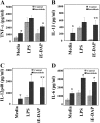
Effect of nucleotide-binding oligomerization domain 1 (NOD)1 and Toll-like receptor 4 (TLR 4) activation on cytokine profiles in cultured bronchoalveolar lavage (BAL) cells. BAL cells from patients with and without sarcoidosis were washed twice with phosphate-buffered saline, counted, and 106 cells were cultured in media containing 1% serum. Cells were incubated with γ-D-glutamyl-meso-diaminopimelic acid (iE-DAP) (1 μg/ml) or LPS (1 ng/ml) for 16 hours or left without treatment. After 16 hours, cytokines were measured in culture supernatants via ELISA for (A) tumor necrosis factor (TNF)-α, (B) IL-1β, (C) IL12/IL23p40, and (D) IL-6. Data are presented as mean ± SEM of 24 patients and 10 control subjects measured in triplicate. It is important to note that there were differences within each group in terms of response to ligands and cytokine production. Using analysis of variance Mann-Whitney U test P < 0.05 was considered significant. *P < 0.05; **P < 0.001. Significant differences were found for IL-1β and IL12/IL23p40 with iE-DAP stimulation (P < 0.001) between the groups.
Figure 3.
Figure 3.
Nucleotide-binding oligomerization domain 1 (NOD1) and Toll-like receptor 4 (TLR 4) stimulation leads to p38 and extracellular signal–regulated kinase (ERK) mitogen-activated protein (MAP) kinase phosphorylation in cultured human bronchoalveolar lavage (BAL) cells. Whole cell extracts were prepared from freshly obtained cells collected during BAL of control subjects and patients, and 30 μg of proteins were subjected to sodium dodecyl sulfate-polyacrylamide gel electrophoresis and Western blot analysis. (A) Whole cell lysates of four control subjects (C1–C4) and four patients (P1–P4). Blots were probed with antibodies against the phosphorylated forms of ERK and p38 as well as total ERK and total p38. Equal loading was confirmed using α-tubulin antibody. (B) BAL cells obtained from control subjects and patients were placed in culture immediately after isolation with and without γ-D-glutamyl-meso-diaminopimelic acid (iE-DAP) (1 μg/ml) or LPS (1 ng/ml) for 45 minutes. Phosphorylation of p38 in BAL cells of patients and control subjects was analyzed using whole cell lysates and specific antibodies against the phosphorylated form (Thr180/Tyr182) and total p38. Representative results are shown for two patients and two control subjects out of a total of n = 12 in each group. (C) Phosphorylation of ERK in BAL cells of patients and control subjects in response to LPS, iE-DAP, and a combination of both. Western blot analysis was performed using antibodies against the phosphorylated form of ERK (Thr202/Tyr204). Equal loading was determined using total ERK. (D) Densitometric values (mean ± SEM) of phosphorylated form of ERK and p38 of eight different experiments. Using analysis of variance Mann-Whitney U test P < 0.05 was considered significant. *P < 0.05; **P < 0.001. Significant differences were found in phosphorylated form of both p38 and ERK kinase in disease and control subjects.
Figure 4.
Figure 4.
Effect of inhibition of extracellular signal–regulated kinase (ERK) and p38 phosphorylation on tumor necrosis factor (TNF)-α and IL-12p40 production in bronchoalveolar lavage (BAL) cells of patients with sarcoidosis. Cells obtained from patients during BAL were counted and placed in culture immediately after isolation. Cells were pretreated either with (A and C) SB203580 or (B and D) PD98059 30 minutes before treatment with γ-D-glutamyl-meso-diaminopimelic acid (iE-DAP) (1 μg/ml) or LPS (1 ng/ml) for 16 hours. (A and B) TNF-α and (C and D) IL-12/IL-23p40 immunoreactivity in supernatants was analyzed by ELISA. Data presented are mean ± SEM of eight patients analyzed in triplicate. Using analysis of variance Mann-Whitney U test P < 0.05 was considered significant. *P < 0.05; **P < 0.001. (E) SB203580 abolishes p38 phosphorylation but increases ERK phosphorylation in BAL cells. Cells (5 × 10⁷) obtained from patients with sarcoidosis (n = 4) were placed in culture immediately after isolation in the presence or absence of SB203580 30 minutes before treatment with iE-DAP (1 μg/ml) or LPS (1 ng/ml) for 45 minutes or left in media without any treatment (control). Whole cell lysates were subjected to Western blot analysis and immunoblotting using antibodies recognizing the active (phosphorylated) form of ERK and p38. Several treatments with other ligands as well as PD98059 were cut from the original blot and excluded in the final image for clarity of image. Equal loading of proteins was confirmed using total ERK and p38 antibodies. As shown, SB203580 inhibited phosphorylation of p38 while increasing ERK phosphorylation. The results were replicated in six different patients. (F) Densitometric values (mean ± SEM) of phosphorylated form of p38 of four different experiments. Using analysis of variance Mann-Whitney U test P < 0.05 was considered significant. **P < 0.001. Significant differences were found between the densitometric units of pp38 in presence and absence of SB203580.
Figure 5.
Figure 5.
Mitogen-activated protein kinase phosphate (MKP)-1 and MKP-3 expression in bronchoalveolar lavage (BAL) cells and healthy peripheral blood monocytes (PBMCs). (A) MKP-3 is down-regulated in response to nucleotide-binding oligomerization domain–like receptor (NLR) and Toll-like receptor 4 (TLR 4) ligands. BAL cells (5 × 10⁷ cells) obtained from patients with sarcoidosis and control subjects were placed in culture immediately after isolation and treated with γ-D-glutamyl-meso-diaminopimelic acid (iE-DAP) (1 μg/ml) or LPS (1 ng/ml) for 45 minutes or left in media without any treatment (control). Whole cell lysates were subjected to Western blot analysis using antibodies recognizing the active phosphorylated form of extracellular signal–regulated kinase (ERK) and a polyclonal antibody against human MKP-3. Equal loading of protein was confirmed using total ERK or β-actin antibodies. Patients demonstrated higher basal levels of MKP3 compared with control subjects. Both groups responded with a down-regulation of MKP3 after nucleotide-binding oligomerization domain 1 (NOD1) and LPS treatment, with patients responding to a lesser degree. Three other treatments with different ligands were cut from the original blot and excluded from the present image. Representative Western blot analyses from six separate experiments (patients) are shown. (B) PBMCs of healthy volunteers were isolated and cells were cultured to a density of 5 × 10⁷ per well and treated with 100 ng/ml LPS for different periods of time. Total cell lysates from human monocytes (n = 3) were subjected to Western blot analysis using antibodies specific for the phosphorylated form of p38 (Thr180/Tyr182), total p38, and MKP-1. Two lanes (treated with different ligands) from the image for the total p38 and phospho p38 were excluded. (C) Dynamic relation of p38 and MKP-1 in isolated PBMC of healthy subjects. Densitometric quantifications from Western blots were performed from three separate experiments for phospho p38 and MKP-1 in response to LPS. The intensity of bands for phospho p38 and MKP1 are plotted against time. (D) Lack of MKP-1 induction in BAL cells of patients with sarcoidosis. BAL cells (5 × 10⁷ cells) obtained from patients with sarcoidosis (n = 12) were placed in culture and treated with iE-DAP (1 μg/ml) or LPS (1 ng/ml) for 60 minutes or left in media without any treatment (media). Whole cell lysates were subjected to Western blot analyses using antibodies specific to phospho p38 (upper panel), MKP-1, and total p38 (as control). (E) Dexamethasone induces MKP-1 in BAL cells. Cells of patients (n = 3) were pretreated with dexamethasone (100 ng/ml) for 45 minutes or kept in media. The cells were treated with NOD1 or TLR4 ligands as indicated. Immunoblotting was performed using phospho p38 (Thr180/Tyr182), and MKP1 antibodies. Equal loading was determined using total p38 antibody. This is a representative Western blot from three different experiments. (F) BAL cells obtained from patients were counted and placed in culture immediately after isolation. Cells were pretreated either with 100 ng/ml dexamethasone for 45 minutes before stimulation with iE-DAP (1 μg/mL) or LPS (1 ng/mL) for 16 hours. Tumor necrosis factor (TNF)-α immunoreactivity in supernatants was analyzed by ELISA. Using analysis of variance Mann-Whitney U test P < 0.05 was considered significant. *P < 0.05.
Figure 6.
Figure 6.
Adenovirus-mediated mitogen-activated protein kinase phosphate (MKP-1) overexpression in bronchoalveolar lavage (BAL) cells of patients with sarcoidosis abrogates p38 phosphorylation and tumor necrosis factor (TNF)-α and IL-12p40 production in response to Toll-like receptor 4 (TLR 4) and nucleotide-binding oligomerization domain 1 (NOD1) agonists. BAL cells of patients (n = 4) were infected with adenovirus-expressing GFP (AdGFP) or adenoviruses expressing MKP-1 (AdMKP-1) at a multiplicity of infection (MOI) of 400. Twenty-four hours post infection cells were stimulated with γ-D-glutamyl-meso-diaminopimelic acid (iE-DAP) (1 μg/ml) or LPS (1 ng/ml) for 45 minutes. Immunoblotting was performed using phospho-p38 (Thr180/Tyr182), phospho-extracellular signal–regulated kinase (ERK) (Thr202/Tyr204), and MKP-1 antibodies. (A) Equal loading was evaluated using total p38 or total ERK antibodies. Different MOI of AdMKP1 were used for the experiments; in the present figure some lanes representing the MOI of 200 were excluded (spliced out). Representative Western blot is shown from four separate experiments. Infected BAL cells were treated with iE-DAP (1 μg/ml) or LPS (1 ng/ml) or kept in media for 16 hours. After 16 hours cytokines were measured in culture supernatants via ELISA for (B) TNF-α and (C) IL12/IL23p40. Data are presented as mean ± SEM of four patients. Using analysis of variance Mann-Whitney U test P < 0.05 was considered significant. *P < 0.05; **P < 0.001.

Comment in

Similar articles

Cited by

References

    1. Statement on sarcoidosis. Joint statement of the American Thoracic Society (ATS), the European Respiratory Society (ERS) and the World Association of Sarcoidosis and Other Granulomatous Disorders (WASOG) adopted by the ATS Board of Directors and by the ERS Executive Committee, February 1999. Am J Respir Crit Care Med 1999;160:736–755. - PubMed
    1. Mathew S, Bauer KL, Fischoeder A, Bhardwaj N, Oliver SJ. The anergic state in sarcoidosis is associated with diminished dendritic cell function. J Immunol 2008;181:746–755. - PMC - PubMed
    1. Prior C, Knight RA, Herold M, Ott G, Spiteri MA. Pulmonary sarcoidosis: patterns of cytokine release in vitro. Eur Respir J 1996;9:47–53. - PubMed
    1. Chen ES, Wahlstrom J, Song Z, Willett MH, Wiken M, Yung RC, West EE, McDyer JF, Zhang Y, Eklund A, et al. T cell responses to mycobacterial catalase-peroxidase profile a pathogenic antigen in systemic sarcoidosis. J Immunol 2008;181:8784–8796. - PMC - PubMed
    1. Furukawa A, Uchida K, Ishige Y, Ishige I, Kobayashi I, Takemura T, Yokoyama T, Iwai K, Watanabe K, Shimizu S, et al. Characterization of propionibacterium acnes isolates from sarcoid and non-sarcoid tissues with special reference to cell invasiveness, serotype, and trigger factor gene polymorphism. Microb Pathog 2009;46:80–87. - PubMed

Publication types

MeSH terms

Substances