. 2010 Nov;76(22):7491-9.
doi: 10.1128/AEM.01614-10. Epub 2010 Sep 17.

New insights into the fructosyltransferase activity of Schwanniomyces occidentalis ß-fructofuranosidase, emerging from nonconventional codon usage and directed mutation

Affiliations

New insights into the fructosyltransferase activity of Schwanniomyces occidentalis ß-fructofuranosidase, emerging from nonconventional codon usage and directed mutation

Miguel Alvaro-Benito et al. Appl Environ Microbiol. 2010 Nov.

Abstract

Schwanniomyces occidentalis β-fructofuranosidase (Ffase) releases β-fructose from the nonreducing ends of β-fructans and synthesizes 6-kestose and 1-kestose, both considered prebiotic fructooligosaccharides. Analyzing the amino acid sequence of this protein revealed that it includes a serine instead of a leucine at position 196, caused by a nonuniversal decoding of the unique mRNA leucine codon CUG. Substitution of leucine for Ser196 dramatically lowers the apparent catalytic efficiency (k(cat)/K(m)) of the enzyme (approximately 1,000-fold), but surprisingly, its transferase activity is enhanced by almost 3-fold, as is the enzymes' specificity for 6-kestose synthesis. The influence of 6 Ffase residues on enzyme activity was analyzed on both the Leu196/Ser196 backgrounds (Trp47, Asn49, Asn52, Ser111, Lys181, and Pro232). Only N52S and P232V mutations improved the transferase activity of the wild-type enzyme (about 1.6-fold). Modeling the transfructosylation products into the active site, in combination with an analysis of the kinetics and transfructosylation reactions, defined a new region responsible for the transferase specificity of the enzyme.


Figures

FIG. 1.
FIG. 1.
(A) Alignment of the regions showing differences between the β-fructofuranosidase from S. occidentalis described here (Ffase) and that previously reported (P24133). The differences are indicated in boldface. The Glu230 involved in sucrose hydrolysis, which is not present in P24133, is marked with an asterisk, and the Ser196 modified to Leu196 in S. cerevisiae is underlined. The superscript numbers indicate the amino acid positions. (B) Ffase was purified from S. occidentalis and digested with trypsin. The sequence of the peptide of 2,005 m/z identified by MALDI-TOF and generated by microspray-ion trap MS analysis is shown.
FIG. 2.
FIG. 2.
Analysis of the enzymes expressed in S. occidentalis (wild type) or in S. cerevisiae (Ffase-Ser196 and Ffase-Leu196). (A) Zymogram (a), Western blot (b), and purification of proteins (c) from S. cerevisiae expressing Ffase-Ser196 (lanes 1) or Ffase-Leu196 (lanes 2) and S. occidentalis (lanes 3). The hydrolase activity of 10 μg of total extracellular proteins was revealed in situ using sucrose as the substrate (a), and 1 μg of each protein was immunoblotted and probed with anti-INV antibodies (b). The purified proteins (10 μg) were subjected to SDS-PAGE and Coomassie stained (c). The numbers on the left (a and b) indicate the positions of molecular mass standards in kDa, and the numbers on the right (c) indicate the molecular masses assigned to the Ffase under the different conditions assayed. (B) Time course of FOS production catalyzed by the Ffase enzymes expressed in S. occidentalis (triangles) and in S. cerevisiae containing Ser (squares) or Leu (rhombus) at position 196. The data are represented as the percentage of FOS (wt/wt) in the total sugar composition of the reaction mixture. The total reaction volume was 2 ml, and 0.3 U of purified enzyme in 0.2 M sodium acetate buffer, pH 5.6, was used. The reaction temperature was 50°C for the wild-type and Ffase-Ser196 enzymes and 45°C for the Ffase-Leu196 variant. (C) HPLC chromatogram corresponding to the reactions of maximum FOS production obtained with the Ffase-Ser196 (6 h) and Ffase-Leu196 (72 h) expressed in S. cerevisiae. The detector response scales for both chromatograms were the same: 1, fructose; 2, glucose; 3, sucrose; 4, 1-kestose; and 5, 6-kestose. (D) Maximum FOS and 6-kestose concentrations (g liter−1) produced by the wild-type (wt), Ffase-Ser196 (Ser196), and Ffase-Leu196 (Leu196) enzymes.
FIG. 3.
FIG. 3.
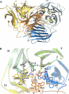
(A) Cartoon of the three-dimensional structure of the S. occidentalis Ffase. Two monomers (blue and orange) associate to form a tight dimer through mainly polar and hydrophobic interactions. The two domains within each monomer are shaded distinctly. The residues investigated in this work are represented as sticks. (B) The catalytic domain folds into a propeller made up of five blades (I to V), each represented in a different color. The residues mentioned in the text are shown as sticks with the same color code. Fructose in the Ffase active site is represented in grey. Asp50 (NDPNG), Asp179 (RDP), and Glu230 (EC) are the catalytic residues. The putative position of a modeled Leu196 is represented in grey.
FIG. 4.
FIG. 4.
(A) Multiple alignments of Ffase and several GH32 proteins in the regions surrounding the sites mutated. The accession code and organism source are indicated in the first column. Identical residues are shaded in black, whereas conserved and semiconserved residues are shaded in dark and light grey, respectively. I, β-fructofuranosidase, EC 3.2.1.26; II, β-fructosidase, EC 3.2.1.80; III fructosyltransferases 2.4.1 (99/100/243); K. lactis, Kluyveromyces lactis; D. hansenii, Debaryomyces hansenii; P. jardinii, Pichia jardinii; S. pombe, Schizosaccharomyces pombe; T. aestivum, Triticum aestivum; Z. maydis, Zea maydis; O. sativa, Oryza sativa; V. faba, Vicia faba; A. cepa, Allium cepa; H. vulgare, Hordeum vulgare; P. sativum, Pisum sativum; K. marxianus, Kluyveromyces marxianus; A. fumigatus, Aspergillus fumigatus; H. tuberosus, Helianthus tuberosus; V. discolor, Viguiera discolor; C. scolymus, Cichorium scolymus; A. officinalis, Asparagus officinalis. (B) Amino acid changes included in the Leu or Ser196 background.
FIG. 5.
FIG. 5.
FOS produced by Ffase-Ser196 and Ffase-Leu196 mutants. The reactions were carried out on sucrose. The concentrations of the total FOS (light grey) and 6-kestose (dark grey) obtained are indicated.
FIG. 6.
FIG. 6.
(A) Close-up view of the active site in S. occidentalis Ffase in a complex with fructose (PDB code 3KF3). Putative positions for 1-kestose (green; left) and 6-kestose (yellow; right) transfructosylation products were modeled into the active site of Ffase. (B and C) The fructan 1-exohydrolase IIa from C. intybus (CiFEH) complexed with 1-kestose (PDB code 2AEZ) (B) and the invertase from T. maritima complexed with raffinose (PDB code 1W2T) (C). The ligands found in the crystals are represented as white sticks. As can be seen in panel A, the active site of Ffase (blue) is also shaped by the adjacent subunit (orange), and it defines subsites in addition to those found in the bacterial and plant enzymes.

Similar articles

Cited by

References

    1. Abarca, D., M. Fernández-Lobato, L. del Pozo, and A. Jiménez. 1991. Isolation of a new gene (SWA2) encoding an alpha amylase from Schwanniomyces occidentalis and its expression in Saccharomyces cerevisiae. FEBS Lett. 279:41-44. - PubMed
    1. Alberto, F., C. Bignon, G. Sulzenbacher, B. Henrissat, and M. Czjzek. 2004. The three-dimenisonal structure of invertase (β-fructosidase) from Thermotoga maritima reveals a bimodular arrangement and an evolutionary relationship between retaining and inverting glycosidases. J. Biol. Chem. 279:18903-18910. - PubMed
    1. Alberto, F., E. Jordi, B. Henrissat, and M. Czjzek. 2006. Crystal structure of inactivated Thermotoga maritima invertase in complex with the trisaccharide substrate raffinose. Biochem. J. 395:457-462. - PMC - PubMed
    1. Altenbach, D., E. Rudiño-Pineira, C. Olvera, T. Boller, A. Wiemken, and T. Ritsema. 2009. An acceptor-substrate binding site determining glycosyl transfer emerges from mutant analysis of a plant vacuolar invertase and a fructosyltransferase. Plant Mol. Biol. 69:47-56. - PMC - PubMed
    1. Álvaro-Benito, M., M. de Abreu, L. Fernández-Arrojo, F. J. Plou, J. Jiménez-Barbero, A. Ballesteros, J. Polaina, and M. Fernández-Lobato. 2007. Characterization of a β-fructofuranosidase from Schwanniomyces occidentalis with transfructosylating activity yielding the prebiotic 6-kestose. J. Biotechnol. 132:75-81. - PubMed

Publication types

Associated data

LinkOut - more resources