. 2010 Nov;9(11):1661-8.
doi: 10.1128/EC.00166-10. Epub 2010 Sep 17.

The antiretroviral lectin cyanovirin-N targets well-known and novel targets on the surface of Entamoeba histolytica trophozoites

Affiliations

The antiretroviral lectin cyanovirin-N targets well-known and novel targets on the surface of Entamoeba histolytica trophozoites

Andrea Carpentieri et al. Eukaryot Cell. 2010 Nov.

Abstract

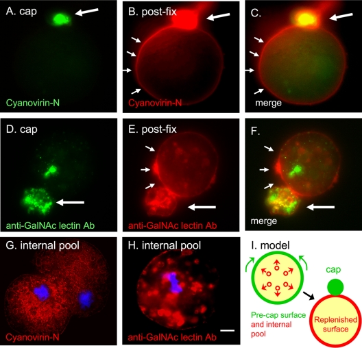
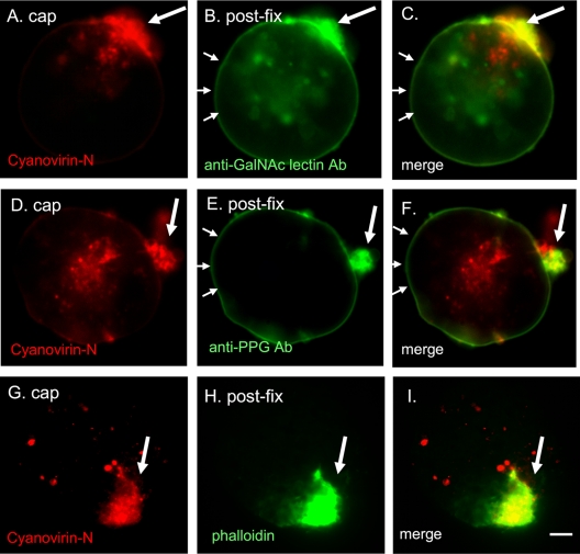
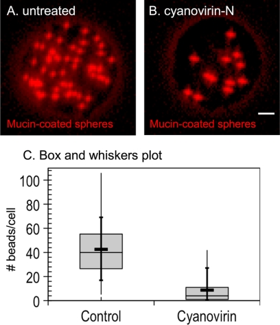
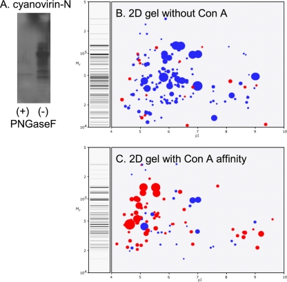
Entamoeba histolytica, the protist that causes amebic dysentery and liver abscess, has a truncated Asn-linked glycan (N-glycan) precursor composed of seven sugars (Man(5)GlcNAc(2)). Here, we show that glycoproteins with unmodified N-glycans are aggregated and capped on the surface of E. histolytica trophozoites by the antiretroviral lectin cyanovirin-N and then replenished from large intracellular pools. Cyanovirin-N cocaps the Gal/GalNAc adherence lectin, as well as glycoproteins containing O-phosphodiester-linked glycans recognized by an anti-proteophosphoglycan monoclonal antibody. Cyanovirin-N inhibits phagocytosis by E. histolytica trophozoites of mucin-coated beads, a surrogate assay for amebic virulence. For technical reasons, we used the plant lectin concanavalin A rather than cyanovirin-N to enrich secreted and membrane proteins for mass spectrometric identification. E. histolytica glycoproteins with occupied N-glycan sites include Gal/GalNAc lectins, proteases, and 17 previously hypothetical proteins. The latter glycoproteins, as well as 50 previously hypothetical proteins enriched by concanavalin A, may be vaccine targets as they are abundant and unique. In summary, the antiretroviral lectin cyanovirin-N binds to well-known and novel targets on the surface of E. histolytica that are rapidly replenished from large intracellular pools.


Figures

Fig. 1.
Fig. 1.
Capped cyanovirin-N-binding glycoproteins and Gal/GalNAc lectins are rapidly replenished on the E. histolytica surface from large intracellular pools. (A) Cyanovirin-N forms a tight green cap (large arrow) on the surface of trophozoites warmed for 15 min prior to fixation. (B) The same organism was fixed and then labeled with red cyanovirin-N. Cyanovirin-N binding sites that are replenished on the E. histolytica surface are marked with small arrows. (D) Similar results were obtained with a monoclonal antibody to the Gal/GalNAc lectin, which forms a tight cap on trophozoites prior to fixation (green). (E) After fixation, the Gal/GalNAc lectin that is replenished on the parasite surface is shown with a red anti-Gal/GalNAc antibody. Merged panels are as indicated. (G) Cyanovirin-N (red) binds to a large interior pool of glycoproteins in a fixed and permeabilized ameba. (H) Similar results were obtained with antibodies to the Gal/GalNAc lectin. In panels G and H nuclei are stained with DAPI (blue). Bar, 5 μm. (I) A model for the redistribution of glycoproteins binding cyanovirin-N or anti-GalNAc antibodies from large intracellular pools during capping. See Fig. S1 in the supplemental material for images of cyanovirin-N and anti-Gal/GalNAc antibody labeling of uncapped parasites. Ab, antibody.
Fig. 2.
Fig. 2.
Cyanovirin-N cocaps and partially depletes the Gal/GalNAc lectin and PPG. E. histolytica trophozoites were capped with cyanovirin-N (red), fixed, and then labeled with monoclonal antibodies to the Gal/GalNAc lectin (green) or to PPG (green). In each case, cyanovirin-N is present in a relatively tight cap (single large arrow), while the Gal/GalNAc lectin and PPG are each present in the cap and along the surface of the protist (series of small arrows). A cyanovirin-N-induced cap on the surface of an E. histolytica trophozoite (G) colocalizes with phalloidin (H) that binds filamentous actin. Merged panels are as indicated. Bar, 5 μm. Ab, antibody.
Fig. 3.
Fig. 3.
Cyanovirin-N inhibits phagocytosis of mucin-coated spheres by E. histolytica trophozoites. Fluorescence micrograph of a control E. histolytica trophozoite (A) that phagocytoses many mucin-coated spheres (red). In contrast, a representative cyanovirin-N-treated E. histolytica trophozoite (B) phagocytoses many fewer red spheres. Bar, 5 μm. (C) Phagocytosis of mucin-coated beads, as illustrated by a modified box and whiskers plot, in which the means are marked with a heavy horizontal line and the medians are marked by a light horizontal line. The lower and upper medians are the edges of each gray box, while the vertical bar shows the minimum and maximum values and the 10th and 90th percentiles (crosses).
Fig. 4.
Fig. 4.
E. histolytica secreted and membrane proteins are markedly enriched by a ConA affinity column. (A) Cyanovirin-N binding to a Western blot of ConA-enriched E. histolytica proteins is removed by prior treatment with PNGaseF. Computer-derived, two-dimensional (2D) protein gels show mass spectrometry data from a representative experiment where unfractionated E. histolytica trophozoite proteins (B) and trophozoite proteins after ConA affinity (C) were identified. The size of each spot is proportional to the peptide coverage of the protein. Secreted proteins, which are markedly enriched after ConA, are shown in red. Nucleocytosolic proteins, which are abundant in unfractionated proteins, are shown in blue.

Similar articles

Cited by

References

    1. Adams E. W., Ratner D. M., Bokesch H. R., McMahon J. B., O'Keefe B. R., Seeberger P. H. 2004. Oligosaccharide and glycoprotein microarrays as tools in HIV glycobiology; glycan-dependent gp120/protein interactions. Chem. Biol. 11:875–881 - PubMed
    1. Altschul S. F., Madden T. L., Schaffer A. A., Zhang J., Zhang Z., Miller W., Lipman D. J. 1997. Gapped BLAST and PSI-BLAST: a new generation of protein database search programs. Nucleic Acids Res. 25:3389–3402 - PMC - PubMed
    1. Arhets P., Gounon P., Sansonetti P., Guillen N. 1995. Myosin II is involved in capping and uroid formation in the human pathogen Entamoeba histolytica. Infect. Immun. 63:4358–4367 - PMC - PubMed
    1. Aurrecoechea C., Brestelli J., Brunk B. P., Fischer S., Gajria B., Gao X., et al. 2010. EuPathDB: a portal to eukaryotic pathogen databases. Nucleic Acids Res. 38:D415–419 - PMC - PubMed
    1. Banerjee S., Vishwanath P., Cui J., Kelleher D. J., Gilmore R., Robbins P. W., Samuelson J. 2007. Evolution of quality control of protein-folding in the ER lumen. Proc. Natl. Acad. Sci. U. S. A. 104:11676–11681 - PMC - PubMed

Publication types

MeSH terms