

Activation of NFATc1 is directly mediated by IP3 in adult cardiac myocytes
- PMID: 20852052
- PMCID: PMC2993207
- DOI: 10.1152/ajpheart.00470.2010
Activation of NFATc1 is directly mediated by IP3 in adult cardiac myocytes
Abstract
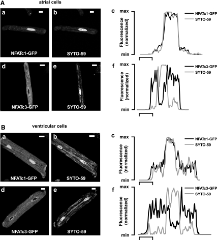
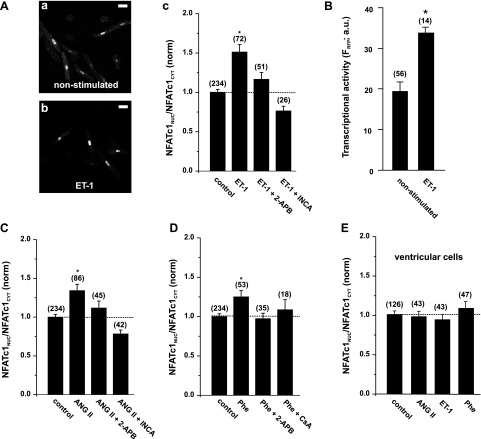
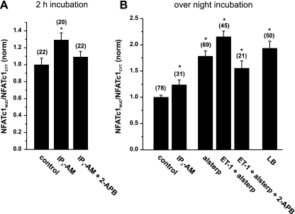
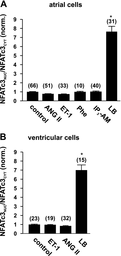
The Ca(2+)-sensitive nuclear factor of activated T cell (NFAT) transcription factors are implicated in cardiac development and cellular remodeling associated with cardiac disease. In adult myocytes it is not resolved what specific Ca(2+) signals control the activity of different NFAT isoforms in an environment that undergoes large changes of intracellular Ca(2+) concentration with every heart beat. Cardiac myocytes possess the complete inositol 1,4,5-trisphosphate (IP(3))/Ca(2+)-signaling cassette; however, its physiological and pathological significance has been a matter of ongoing debate. Therefore, we tested the hypothesis whether IP(3) receptor activation regulates NFAT activity in cardiac myocytes. We used confocal microscopy to quantify the nuclear localization of NFATc1-green fluorescent protein (GFP) and NFATc3-GFP fusion proteins (quantified as the ratio of nuclear NFAT to cytoplasmic NFAT) in response to stimulation with neurohumoral agonists. In rabbit atrial myocytes, an overnight stimulation with endothelin-1, angiotensin II, and phenylephrine induced nuclear accumulation of NFATc1 that was sensitive to calcineurin inhibitors (cyclosporin A or inhibitor of NFAT-calcineurin association-6) and prevented by the IP(3) receptor inhibitor 2-aminoethoxydiphenyl borate. Furthermore, a direct elevation of intracellular IP(3) with a cell-permeable IP(3) acetoxymethyl ester (10 μM) induced nuclear localization of NFATc1. With a fluorescence-based in vivo assay, we showed that endothelin-1 also enhanced the transcriptional activity of NFATc1 in atrial cells. The agonists failed to activate NFATc1 in rabbit ventricular cells, which express IP(3) receptors at a lower density than atrial cells. They also did not activate NFATc3, an isoform that is highly influenced by nuclear export processes, in both cell types. Our data show that the second messenger IP(3) is directly involved in the activation of NFATc1 in adult atrial cardiomyocytes.
Figures
Similar articles
-
Isoform- and tissue-specific regulation of the Ca(2+)-sensitive transcription factor NFAT in cardiac myocytes and heart failure.Am J Physiol Heart Circ Physiol. 2010 Jun;298(6):H2001-9. doi: 10.1152/ajpheart.01072.2009. Epub 2010 Mar 19. Am J Physiol Heart Circ Physiol. 2010. PMID: 20304816 Free PMC article.
-
NFAT transcription factor regulation by urocortin II in cardiac myocytes and heart failure.Am J Physiol Heart Circ Physiol. 2014 Mar;306(6):H856-66. doi: 10.1152/ajpheart.00353.2013. Epub 2014 Jan 17. Am J Physiol Heart Circ Physiol. 2014. PMID: 24441548 Free PMC article.
-
Angiotensin II and norepinephrine activate specific calcineurin-dependent NFAT transcription factor isoforms in cardiomyocytes.J Appl Physiol (1985). 2011 Nov;111(5):1278-89. doi: 10.1152/japplphysiol.01383.2010. Epub 2011 Apr 7. J Appl Physiol (1985). 2011. PMID: 21474694
-
Chromogranins and inositol 1,4,5-trisphosphate-dependent Ca(2+)-signaling in cardiomyopathy and heart failure.Curr Med Chem. 2012;19(24):4068-73. doi: 10.2174/092986712802430045. Curr Med Chem. 2012. PMID: 22834797 Review.
-
Emerging roles of inositol 1,4,5-trisphosphate signaling in cardiac myocytes.J Mol Cell Cardiol. 2008 Aug;45(2):128-47. doi: 10.1016/j.yjmcc.2008.05.014. Epub 2008 Jun 15. J Mol Cell Cardiol. 2008. PMID: 18603259 Free PMC article. Review.
Cited by
-
Left Atrial Myocardium in Arterial Hypertension.Cells. 2022 Oct 8;11(19):3157. doi: 10.3390/cells11193157. Cells. 2022. PMID: 36231118 Free PMC article. Review.
-
Inositol 1,4,5-trisphosphate receptors in cardiomyocyte physiology and disease.Philos Trans R Soc Lond B Biol Sci. 2022 Nov 21;377(1864):20210319. doi: 10.1098/rstb.2021.0319. Epub 2022 Oct 3. Philos Trans R Soc Lond B Biol Sci. 2022. PMID: 36189803 Free PMC article. Review.
-
Cytosolic and nuclear calcium signaling in atrial myocytes: IP3-mediated calcium release and the role of mitochondria.Channels (Austin). 2015;9(3):129-38. doi: 10.1080/19336950.2015.1040966. Channels (Austin). 2015. PMID: 25891132 Free PMC article.
-
miR-155 functions downstream of angiotensin II receptor subtype 1 and calcineurin to regulate cardiac hypertrophy.Exp Ther Med. 2016 Sep;12(3):1556-1562. doi: 10.3892/etm.2016.3506. Epub 2016 Jul 5. Exp Ther Med. 2016. PMID: 27588076 Free PMC article.
-
Inositol 1,4,5-trisphosphate receptor - reactive oxygen signaling domain regulates excitation-contraction coupling in atrial myocytes.J Mol Cell Cardiol. 2022 Feb;163:147-155. doi: 10.1016/j.yjmcc.2021.10.006. Epub 2021 Oct 28. J Mol Cell Cardiol. 2022. PMID: 34755642 Free PMC article.
References
-
- Bers DM. Calcium cycling and signaling in cardiac myocytes. Annu Rev Physiol 70: 23–49, 2008. - PubMed
-
- Crabtree GR, Olson EN. NFAT signaling: choreographing the social lives of cells. Cell 109, Suppl: S67–S79, 2002 - PubMed
-
- de la Pompa JL, Timmerman LA, Takimoto H, Yoshida H, Elia AJ, Samper E, Potter J, Wakeham A, Marengere L, Langille BL, Crabtree GR, Mak TW. Role of the NF-ATc transcription factor in morphogenesis of cardiac valves and septum. Nature 392: 182–186, 1998 - PubMed
-
- Dolmetsch RE, Lewis RS, Goodnow CC, Healy JI. Differential activation of transcription factors induced by Ca2+ response amplitude and duration. Nature 386: 855–858, 1997 - PubMed
Publication types
MeSH terms
Substances
Grants and funding
LinkOut - more resources
Full Text Sources
Miscellaneous