. 2010 Dec 16;116(25):5455-64.
doi: 10.1182/blood-2010-05-286328. Epub 2010 Sep 17.

Deletion-based mechanisms of Notch1 activation in T-ALL: key roles for RAG recombinase and a conserved internal translational start site in Notch1

Affiliations

Deletion-based mechanisms of Notch1 activation in T-ALL: key roles for RAG recombinase and a conserved internal translational start site in Notch1

Todd D Ashworth et al. Blood. .

Abstract

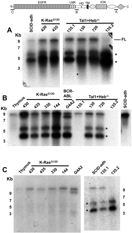
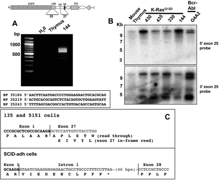
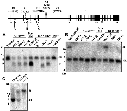
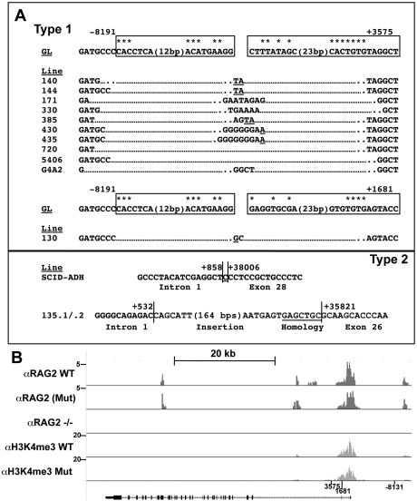
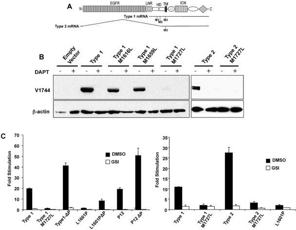
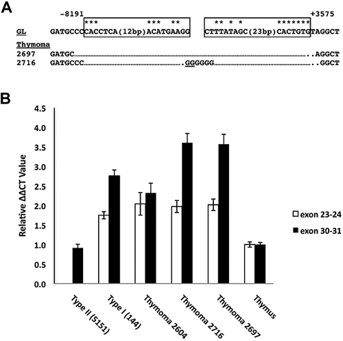
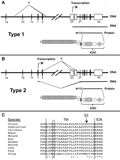
Point mutations that trigger ligand-independent proteolysis of the Notch1 ectodomain occur frequently in human T-cell acute lymphoblastic leukemia (T-ALL) but are rare in murine T-ALL, suggesting that other mechanisms account for Notch1 activation in murine tumors. Here we show that most murine T-ALLs harbor Notch1 deletions that fall into 2 types, both leading to ligand-independent Notch1 activation. Type 1 deletions remove exon 1 and the proximal promoter, appear to be RAG-mediated, and are associated with mRNA transcripts that initiate from 3' regions of Notch1. In line with the RAG dependency of these rearrangements, RAG2 binds to the 5' end of Notch1 in normal thymocytes near the deletion breakpoints. Type 2 deletions remove sequences between exon 1 and exons 26 to 28 of Notch1, appear to be RAG-independent, and are associated with transcripts in which exon 1 is spliced out of frame to 3' Notch1 exons. Translation of both types of transcripts initiates at a conserved methionine residue, M1727, which lies within the Notch1 transmembrane domain. Polypeptides initiating at M1727 insert into membranes and are subject to constitutive cleavage by γ-secretase. Thus, like human T-ALL, murine T-ALL is often associated with acquired mutations that cause ligand-independent Notch1 activation.


Figures

Figure 1
Figure 1
Northern blot analysis of murine T-ALL cell lines reveals 2 types of aberrant Notch1 transcripts. Northern blot analyses used polyA RNAs from normal murine thymus or the indicated cells lines. Panels A, B, and C correspond to Northern blots hybridized to probes A (exon 34), B (exon 26), or C (exon 1), which are homologous to the regions shown in the cartoon of the Notch1 locus above the blots. Numbers correspond to the position of RNA size markers in kilobases. FL indicates full-length Notch1 transcript. *Short Notch1 transcripts.
Figure 2
Figure 2
Structure of type 1 and type 2 Notch1 transcripts. (A) Products amplified from 144 cells, a line expressing type 1 Notch1 transcripts, using a 3′ exon 27 primer and 5′ linker primer, are shown after agarose gel electrophoresis. Sequences of 5′RACE products are shown below; the basepair numbering is in relationship to the A residue in the ATG start site in exon 1 of the Notch1 gene, which is designated position 1. (B) Northern blot analysis of lines expressing type 1 Notch1 transcripts with probes from the 3′ end of exon 25 and the 5′ end of exon 25. The diagram shows the regions of the Notch1 that are encoded by exons 25 and 27, respectively. (C) Sequences of RT-PCR products obtained from murine T-ALL cell lines expressing type II transcripts. Sequencing of products obtained from 130.1 and 130.2 cells revealed a transcript in which exon 1 is spliced out of frame to exon 27. In SCID-adh cells, a product was obtained in which exon 1 is spliced to an intron 1 sequence that contains an in-frame stop codon, which is in turned joined to an internal sequence within exon 28.
Figure 3
Figure 3
Southern blot analysis reveals 2 types of 5′ Notch1 genomic rearrangements in murine cell lines expressing type 1 (I) or type 2 (II) aberrant transcripts. Genomic DNA (10 μg) from the indicated cell lines was digested with Eco R1 and analyzed on Southern blots. Panels A, B, and C correspond to Southern blots hybridized to probes A, B, and C, which are homologous to the regions shown in the diagram of the Notch1 locus above the blots. The positions of genomic Eco R1 sites (R1) around the Notch1 locus are expressed relative to the position of the ATG start codon in exon 1, which is designated position 1. The positions of murine-specific alternative 5′ Notch1 exons 1a, 1b, and 1c are also shown. R indicates rearranged; and GL, normal genomic locus.
Figure 4
Figure 4
Evidence of RAG involvement in Notch1 rearrangements in murine T-ALL. (A) Rearrangements in Notch1 deduced from sequencing of PCR products generated from 11 cell lines with type 1 deletions (top) and 3 cell lines with type 2 deletions (bottom). GL is the sequence of the germline DNA flanking the breakpoints. Nucleotide positions are expressed relative to the ATG start codon in exon 1 of Notch1. Flanking sequences resembling RAG recognition sequences are boxed. Residues matching the consensus RAG signal sequence (a CACATGT heptamer followed by a 12- or 23-bp spacer and the nonameric sequence ACAAAAAAC) are denoted with an asterisk. N nucleotides and P nucleotides (underlined) are also shown. The point of joining in SCID-adh contains a single cytosine residue (underlined) of unknown origin. (B) Distribution of RAG2 binding and H3K4 trimethylation across the murine Notch1 locus. ChIP-Seq was performed with antibodies specific for RAG2 and H3K4-me3 on DNA immunoprecipitated from normal thymocytes (αRAG2 WT), thymocytes expressing a RAG1 D708A mutant (αRAG2 Mut) that binds chromatin but is catalytically inactive, and homozygous RAG2 knockout thymoctyes (αRAG2−/−). Histograms showing sequence reads that aligned to the murine genome are superimposed on a diagram of the Notch1 locus. The y-axis of each histogram corresponds to the number of aligned reads per 106 total reads.
Figure 5
Figure 5
M1727 serves as the translational start site in aberrant type 1 and type 2 Notch1 transcripts. (A) Schematic showing the positions of codons M1616 (M1), M1659 (M2), and M1727 (M3) within type 1 and type 2 Notch1 transcripts relative to the full-length Notch1 polypeptide. (B) Western blot analysis of 293 cells transiently transfected with empty pcDNA3, type 1 transcript cDNA (corresponding to nucleotides 4438-7665 of murine Notch1), type 1 transcript cDNAs bearing M to L point mutations in codons 1616, 1659, or 1727, type 2 transcript cDNA from SCID.adh cells, or type 2 transcript cDNA with an M to L point mutation in codon 1727. Blots were stained with antibodies specific for activated Notch1 (V1744 antibody, Cell Signaling Technology) or β-actin. (C) Notch reporter gene studies conducted in U2OS cells transiently transfected in triplicate with equivalent amounts of a CSLx4 firefly luciferase reporter gene, an internal Renilla luciferase control gene, and empty pcDNA3 vector or pcDNA3 vectors containing the indicated cDNAs. The left-hand panel compares the activities of a murine type 1 Notch1 transcript; a murine type 1 Notch1 transcript containing a M1727L mutation; a murine type 1 Notch1 transcript containing a stop mutation introduced at codon 2415 (ΔP) that deletes the C-terminal 116 amino acids of murine Notch1; a full-length human NOTCH1 cDNA containing a weakly activating NRR mutation, L1601P, with and without a ΔP frameshift mutation at codon 2473 that deletes the C-terminal 83 amino acids of human Notch1; and a full-length human NOTCH1 cDNA containing a strongly activating in-frame insertion in the NRR designated P12, with and without a ΔP frameshift mutation at codon 2473. The right-hand panel compares the activities of murine type 1 and type 2 transcripts with a human NOTCH1 cDNA bearing a L1601P deletion. Each cDNA was tested in the presence and absence vehicle (dimethyl sulfoxide [DMSO]) or vehicle plus the GSI DAPT (10μM). After normalization to an internal Renilla luciferase control, firefly luciferase activities were expressed relative to the empty vector control, which was arbitrarily set to 1. Error bars represent SD. The results shown are representative of 2 independent experiments.
Figure 6
Figure 6
Detection of 5′ deletions and aberrant Notch1 transcripts in primary murine “thymomas.” (A) Sequences of PCR products obtained by amplification of genomic DNA isolated from 2 thymic lymphomas with primers flanking the most common breakpoints associated with type 1 aberrant transcripts. Sites of DNA breakage and joining, as deduced from sequencing of PCR products, are shown. Residues matching the consensus RAG recognition sequence (CACAGTG followed by a 12- or 23-bp spacer and the sequence ACAAAAAAC) are denoted with an asterisk. N nucleotides and P nucleotides (underlined) are also shown. GL indicates germline DNA flanking the breakpoints. Boxes represent sequences resembling RAG signal sequences. (B) Ratiometric Notch1 quantitative RT-PCR analysis. The relative amounts of transcripts containing 5′ (exons 23 and 24) and 3′ (exons 30 and 31) Notch1 sequences were determined for the tumors in panel A and normal murine thymus, a cell line with a homozygous type 2 deletion (135.2), and a cell line with a heterozygous type 1 deletion (144). Each determination was made in triplicate. The results shown are representative of 2 independent experiments.
Figure 7
Figure 7
Mechanisms of ligand-independent ICN1 production in T-ALLs bearing Notch1 deletions. (A-B) Structure and functional consequences of type 1 and type 2 Notch1 deletions. (C) Conservation of M1727 in vertebrate Notch1 receptors. *Identical residues. “:” indicates conserved residues. TM indicates transmembrane domain; S3, site of intramembranous γ-secretase cleavage; and ICN, intracellular Notch.

Comment in

References

    1. Aster JC, Pear WS, Blacklow SC. Notch signaling in leukemia. Annu Rev Pathol. 2008;3:587–613. - PMC - PubMed
    1. Weng AP, Ferrando AA, Lee W, et al. Activating mutations of NOTCH1 in human T cell acute lymphoblastic leukemia. Science. 2004;306(5694):269–271. - PubMed
    1. Gordon WR, Vardar-Ulu D, Histen G, Sanchez-Irizarry C, Aster JC, Blacklow SC. Structural basis for autoinhibition of Notch. Nat Struct Mol Biol. 2007;14(4):295–300. - PubMed
    1. Malecki MJ, Sanchez-Irizarry C, Mitchell JL, et al. Leukemia-associated mutations within the NOTCH1 heterodimerization domain fall into at least two distinct mechanistic classes. Mol Cell Biol. 2006;26(12):4642–4651. - PMC - PubMed
    1. Gordon WR, Roy M, Vardar-Ulu D, et al. Structure of the Notch1-negative regulatory region: implications for normal activation and pathogenic signaling in T-ALL. Blood. 2009;113(18):4381–4390. - PMC - PubMed

Publication types

MeSH terms