. 2010 Dec 3;285(49):37976-86.
doi: 10.1074/jbc.M110.174458. Epub 2010 Sep 18.

Chylomicron- and VLDL-derived lipids enter the heart through different pathways: in vivo evidence for receptor- and non-receptor-mediated fatty acid uptake

Affiliations

Chylomicron- and VLDL-derived lipids enter the heart through different pathways: in vivo evidence for receptor- and non-receptor-mediated fatty acid uptake

Kalyani G Bharadwaj et al. J Biol Chem. .

Abstract

Lipids circulate in the blood in association with plasma lipoproteins and enter the tissues either after hydrolysis or as non-hydrolyzable lipid esters. We studied cardiac lipids, lipoprotein lipid uptake, and gene expression in heart-specific lipoprotein lipase (LpL) knock-out (hLpL0), CD36 knock-out (Cd36(-/-)), and double knock-out (hLpL0/Cd36(-/-)-DKO) mice. Loss of either LpL or CD36 led to a significant reduction in heart total fatty acyl-CoA (control, 99.5 ± 3.8; hLpL0, 36.2 ± 3.5; Cd36(-/-), 57.7 ± 5.5 nmol/g, p < 0.05) and an additive effect was observed in the DKO (20.2 ± 1.4 nmol/g, p < 0.05). Myocardial VLDL-triglyceride (TG) uptake was reduced in the hLpL0 (31 ± 6%) and Cd36(-/-) (47 ± 4%) mice with an additive reduction in the DKO (64 ± 5%) compared with control. However, LpL but not CD36 deficiency decreased VLDL-cholesteryl ester uptake. Endogenously labeled mouse chylomicrons were produced by tamoxifen treatment of β-actin-MerCreMer/LpL(flox/flox) mice. Induced loss of LpL increased TG levels >10-fold and reduced HDL by >50%. After injection of these labeled chylomicrons in the different mice, chylomicron TG uptake was reduced by ∼70% and retinyl ester by ∼50% in hLpL0 hearts. Loss of CD36 did not alter either chylomicron TG or retinyl ester uptake. LpL loss did not affect uptake of remnant lipoproteins from ApoE knock-out mice. Our data are consistent with two pathways for fatty acid uptake; a CD36 process for VLDL-derived fatty acid and a non-CD36 process for chylomicron-derived fatty acid uptake. In addition, our data show that lipolysis is involved in uptake of core lipids from TG-rich lipoproteins.


Figures
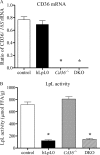
FIGURE 1.
FIGURE 1.
Characterization of LpL/CD36 double knock-out mice. The LpL/CD36 DKO on C57Bl/6J background were generated by a breeding scheme as described under “Experimental Procedures.” 14-Week-old male mice were fasted for 4–5 h before harvesting tissues for all measurements. A, cardiac CD36 mRNA expression normalized to 18S rRNA expression measured by real time RT-PCR. B, cardiac LpL enzyme activity per g wet weight of heart. n = 4–6 per group; *, significantly different from control, p < 0.05.
FIGURE 2.
FIGURE 2.
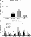
Cardiac total long chain fatty acyl-CoA (LCCoA) content. 12–13-Week-old male mice were fasted for 4–5 h before harvesting the hearts for measurement of cardiac fatty acyl-CoA content by the LC-MS/MS technique described under “Experimental Procedures.” A, cardiac total LCCoA content. B, distribution of LCCoA species in the heart. n = 5–8 per group; *, significantly different from control; #, significantly different from DKO, p < 0.05.
FIGURE 3.
FIGURE 3.
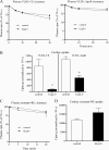
Cardiac VLDL-TG and VLDL-CE uptake. 13–14-Week-old male mice fasted for 4–5 h were administered double-labeled human VLDL particles by tail vein. A, plasma decay curves for VLDL-TG. B, cardiac VLDL-TG uptake 30 min after tracer injection. C, plasma decay curves for VLDL-CE. D, cardiac VLDL-CE uptake 30 min after tracer injection expressed. n = 4–5 per group; *, significantly different from control; #, significantly different from DKO, p < 0.05.
FIGURE 4.
FIGURE 4.
Characterization of plasma lipids in tamoxifen-induced LpL deletion mouse model (LpL−/−). A, representative plasma from control and LpL-depleted mice 2 weeks after tamoxifen injection. B, plasma TG measured 2 weeks after tamoxifen injection. Mice with plasma TG > 400 mg/dL were used to make endogenously labeled CMs in subsequent studies. Lipoprotein TG (C) and TC (D) in control and LpL− /− mice 7 and 35 days after tamoxifen injection. VLDL refers to d < 1.006 fraction that contains both VLDL and CMs. *, significantly different from control, p < 0.05.
FIGURE 5.
FIGURE 5.
Cardiac CM-TG and CM-RE uptake. 10–12-Week-old male mice fasted for 4–5 h were administered double-labeled mouse CMs by tail vein. A, plasma decay curves for CM-TG. B, cardiac CM-TG uptake 15 min after tracer injection. C, plasma decay curves for CM-RE. D, cardiac CM-RE uptake 15 min after tracer injection. n = 5–7 per group; *, significantly different from control, p < 0.05.
FIGURE 6.
FIGURE 6.
Cardiac uptake of VLDL-CE, VLDL-ApoB, and remnants. 12–13-Week-old male mice fasted for 4–5 h were administered 125I-TC-conjugated human VLDL particles along with [3H]CE-labeled VLDL via the tail vein. A, plasma decay curves for VLDL-CE (left panel) and VLDL-ApoB (right panel). B, cardiac uptake 30 min after tracer injection, n = 5–7 per group. 12–14-Week-old male mice fasted for 4–5 h were administered [3H]RE-labeled remnant particles via the tail vein. C, plasma decay curves for ApoE−/− remnant particles. D, cardiac uptake of tracer 20 min after injection, n = 3–5 per group; *, significantly different from control, p < 0.05.
FIGURE 7.
FIGURE 7.
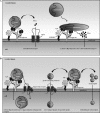
A, schematic model for VLDL- and CM-derived TG uptake pathways. 1) Hydrolysis of VLDL particles generates FFAs that are subsequently transported into cells via the fatty acid transporter CD36 by the facilitated transport process. LpL is shown attached to the cell surface via binding to proteoglycans and its binding partner glycosylphosphatidylinositol-anchored high density lipoprotein-binding protein 1 (GPIHBP1). 2) Lipolysis of TG-rich CM particles generates a higher local concentration of FFAs most of which enter cells by a passive flip-flop and/or non-CD36-mediated mechanism. B, schematic model for LpL-mediated uptake of core lipid. 1) LpL approximates TG-rich lipoproteins (TGRLP) to the cell membrane and increases uptake of particles either via recycling of membranes or along with lipoprotein receptors. 2) LpL creates ApoB-containing remnants that are better ligands for lipoprotein receptors. 3) During lipolysis, apoproteins and lipids, including core lipid esters such as CE and RE, are shed as surface lipids and subsequently transferred to underlying cells.

Similar articles

Cited by

References

    1. Wisneski J. A., Gertz E. W., Neese R. A., Mayr M. (1987) J. Clin. Invest. 79, 359–366 - PMC - PubMed
    1. DeGrella R. F., Light R. J. (1985) Basic Res. Cardiol. 80, Suppl. 2, 107–110 - PubMed
    1. Stremmel W. (1988) J. Clin. Invest. 81, 844–852 - PMC - PubMed
    1. DeGrella R. F., Light R. J. (1980) J. Biol. Chem. 255, 9731–9738 - PubMed
    1. Abumrad N. A., Park J. H., Park C. R. (1984) J. Biol. Chem. 259, 8945–8953 - PubMed

Publication types

MeSH terms