. 2010 Oct;17(10):1202-9.
doi: 10.1038/nsmb.1908. Epub 2010 Sep 19.

Structural basis of HIV-1 resistance to AZT by excision

Affiliations

Structural basis of HIV-1 resistance to AZT by excision

Xiongying Tu et al. Nat Struct Mol Biol. 2010 Oct.

Abstract

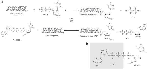
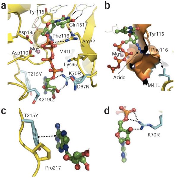
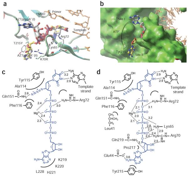
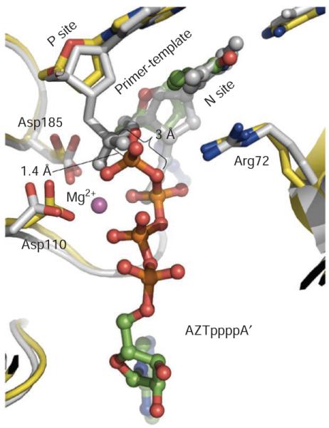
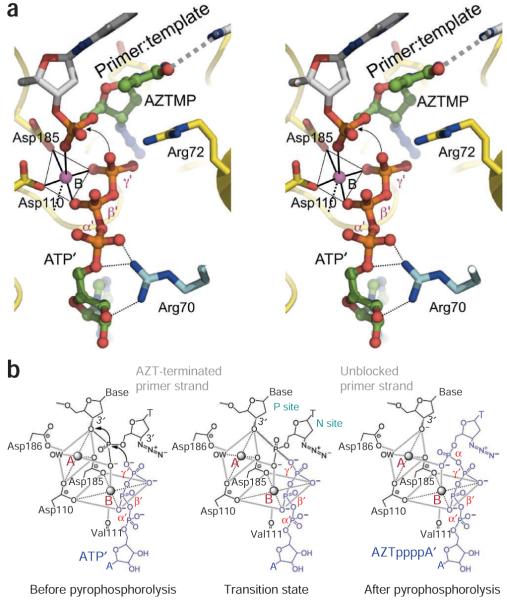
Human immunodeficiency virus (HIV-1) develops resistance to 3'-azido-2',3'-deoxythymidine (AZT, zidovudine) by acquiring mutations in reverse transcriptase that enhance the ATP-mediated excision of AZT monophosphate from the 3' end of the primer. The excision reaction occurs at the dNTP-binding site, uses ATP as a pyrophosphate donor, unblocks the primer terminus and allows reverse transcriptase to continue viral DNA synthesis. The excision product is AZT adenosine dinucleoside tetraphosphate (AZTppppA). We determined five crystal structures: wild-type reverse transcriptase-double-stranded DNA (RT-dsDNA)-AZTppppA; AZT-resistant (AZTr; M41L D67N K70R T215Y K219Q) RT-dsDNA-AZTppppA; AZTr RT-dsDNA terminated with AZT at dNTP- and primer-binding sites; and AZTr apo reverse transcriptase. The AMP part of AZTppppA bound differently to wild-type and AZTr reverse transcriptases, whereas the AZT triphosphate part bound the two enzymes similarly. Thus, the resistance mutations create a high-affinity ATP-binding site. The structure of the site provides an opportunity to design inhibitors of AZT-monophosphate excision.


Figures

Figure 1
Figure 1
Chemical reactions of DNA polymerization and ATP-mediated pyrophosphorolytic excision catalyzed by HIV-1 reverse transcriptase (RT). (a) DNA polymerization incorporates nucleotides and NRTIs into the DNA primer strand (top row). Incorporated NRTIs can be removed by pyrophosphorolysis, a reverse reaction of polymerization, in which the ATP-mediated excision of AZTMP (pAZT) produces AZTppppA and unblocks the DNA primer strand (bottom row, right to left). (b) Chemical structure of AZTppppA. The two-headed nucleotide AZTppppA was chemically synthesized and used for structural studies of excision-product complexes of wild-type and AZTr reverse transcriptases; ATP portion, ATP’; β’ and γ’ phosphates of ATP’ are γ and β phosphates of AZTTP, respectively.
Figure 2
Figure 2
Binding of AZTppppA’ to AZTr HIV-1 reverse transcriptase. (a) Interactions of AZTppppA’ (carbons, green; phosphates, orange) with the mutated residues (cyan) and with the active site residues; hydrogen bonds, dotted lines; nucleic acid, thin lines. (b) Molecular surface representing the azido-binding cleft at the N site. (c) π-π stacking between Tyr215 and the adenine ring, and the hydrophobic interaction between Pro217 and the ribose ring stabilize the binding of ATP’ to AZTr reverse transcriptase. (d) Primary mutation K70R enhances ATP’ binding by interacting with the ribose ring and α phosphate. Structure images were made using PyMOL (http://www.pymol.org/).
Figure 3
Figure 3
AZTppppA’ binds differently to AZTr reverse transcriptase than to wild-type reverse transcriptase. (a) Superposition of the wild-type HIV-1 RT–dsDNA–AZTppppA’ structure (gray) on the AZTr HIV-1 RT–dsDNA–AZTppppA’ structure (cyan). The AMP’ portion of AZTppppA’ binds differently to AZTr reverse transcriptase (yellow) than to wild-type reverse transcriptase (gray). (b) Molecular surface of AZTr reverse transcriptase showing ATP’-binding sites. Site I, where ATP’ binds to wild-type reverse transcriptase, remains almost unchanged in AZTr reverse transcriptase; however, a new ATP’-binding site (site II) is created by AZTr mutations that substantially enhance the binding of ATP’ to AZTr reverse transcriptase. (c) Summary of interactions of AZTppppA’ (blue) with wild-type HIV-1 reverse transcriptase. (d) Summary of interactions of AZTppppA’ with AZTr HIV-1 reverse transcriptase. Hydrogen bonds and metal coordinations, dotted lines. Ribbon and molecular representations were drawn using Schrodinger (http://www.schrodinger.com/).
Figure 4
Figure 4
Superposition of AZTr reverse transcriptase excision product complex (yellow) and AZTr reverse transcriptase–template-primer–AZTMP (N site) complex (gray) structures. AZTppppA’ has green carbon and orange phosphorus atoms. The AZT portions superimpose well; the α-phosphorus atoms are ~1.4 Å apart in the two superimposed structures, indicating the position of the α phosphate before and after AZTTP incorporation; the metal coordination of an α-phosphate oxygen is maintained before and after incorporation of AZTMP. Figure generated using PyMOL.
Figure 5
Figure 5
Mechanism of ATP-mediated excision by HIV-1 reverse transcriptase. (a) Stereo view showing relative locations of DNA-primerterminated AZTMP and the ATP excision substrate; locations based on superposition (Fig. 4) of structures of AZTr RT–dsDNA–AZTppppA’ and AZTr RT–dsDNA (with AZTMP at the primer terminus occupying the N site). The metal A position was not observed in the current structures, therefore only metal B is shown. (b) Scheme for ATP-mediated excision. Catalysis involves two Mg2+ ions, on the basis of our structural results and the reported coordination geometry at the catalytic active site of HIV-1 reverse transcriptase,. The catalytic reactions of polymerization and excision should use the same cations (A and B) in the identical coordination environment. Planes and axes of coordination for both metal ions are gray boxes and lines, respectively. The excision substrate (ATP’) and the product (AZTppppA’) are blue; the prepyrophosphorolysis state was derived by positioning ATP’ in N-site complex structure, and the excision-complex structures represent the post-pyrophosphorolysis state.

Similar articles

Cited by

References

    1. Sarafianos SG, et al. Trapping HIV-1 reverse transcriptase before and after translocation on DNA. J. Biol. Chem. 2003;278:16280–16288. - PubMed
    1. Otwinowski Z, Minor W. Processing of X-ray diffraction data collected in oscillation mode. Methods Enzymol. 1997;276:307–326. - PubMed
    1. Strong M, et al. Toward the structural genomics of complexes: crystal structure of a PE/PPE protein complex from Mycobacterium tuberculosis. Proc. Natl. Acad. Sci. USA. 2006;103:8060–8065. - PMC - PubMed
    1. Collaborative Computational Project, Number 4 The CCP4 suite: programs for protein crystallography. Acta Crystallogr. D Biol. Crystallogr. 1994;50:760–763. - PubMed
    1. Brünger AT, et al. Crystallography & NMR system: A new software suite for macromolecular structure determination. Acta Crystallogr. D Biol. Crystallogr. 1998;54:905–921. - PubMed

MeSH terms