

Loss of microRNAs in thymus perturbs invariant NKT cell development and function
- PMID: 20852654
- PMCID: PMC4002964
- DOI: 10.1038/cmi.2010.49
Loss of microRNAs in thymus perturbs invariant NKT cell development and function
Abstract
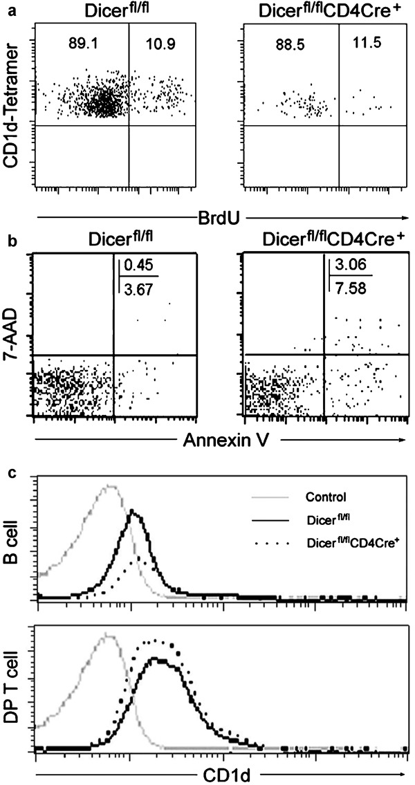
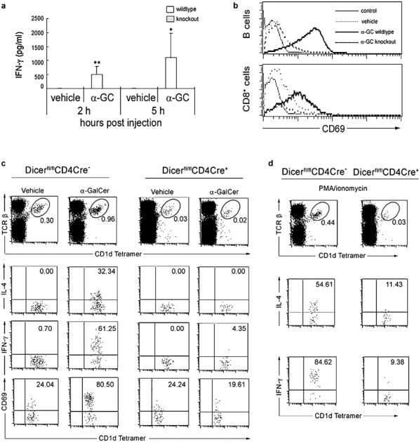
microRNAs (miRNAs) are small noncoding RNAs that mediate RNA interference to suppress protein expression at the translational level. Accumulated evidence indicates that miRNAs play critical roles in various biological processes and disease development, including autoimmune diseases. Invariant natural killer T (iNKT) cells are an unusual CD1d-restricted subset of thymus-derived T cells that are potent regulators of diverse immune responses. Our previous studies with the mouse model of bone marrow-specific Dicer deletion suggest the involvement of Dicer-dependent miRNAs in the development and function of iNKT cells. In the present study, to further dissect the functional levels of Dicer-dependent miRNAs in regulating iNKT cell development, we generated a mouse model with the Dicer deletion in the thymus. Our data indicate that lack of miRNAs following the deletion of Dicer in the thymus severely interrupted the development and maturation of iNKT cells in the thymus and significantly decreased the number of iNKT cells in the peripheral immune organs. miRNA-deficient peripheral iNKT cells display profound defects in activation and cytokine production upon α-galactosylceramide (α-GalCer) stimulation. Our results demonstrate a critical role of the miRNA-dependent pathway in the thymus in the regulation of iNKT cell development and function.
Figures


Similar articles
-
Tie2cre-induced inactivation of the miRNA-processing enzyme Dicer disrupts invariant NKT cell development.Proc Natl Acad Sci U S A. 2009 Jun 23;106(25):10266-71. doi: 10.1073/pnas.0811119106. Epub 2009 Jun 9. Proc Natl Acad Sci U S A. 2009. PMID: 19509335 Free PMC article.
-
Dicer-dependent microRNA pathway controls invariant NKT cell development.J Immunol. 2009 Aug 15;183(4):2506-12. doi: 10.4049/jimmunol.0901361. Epub 2009 Jul 22. J Immunol. 2009. PMID: 19625646
-
Invariant NKT cell development and function in microRNA-223 knockout mice.Int Immunopharmacol. 2011 May;11(5):561-8. doi: 10.1016/j.intimp.2010.11.004. Epub 2010 Nov 19. Int Immunopharmacol. 2011. PMID: 21094288
-
New Genetically Manipulated Mice Provide Insights Into the Development and Physiological Functions of Invariant Natural Killer T Cells.Front Immunol. 2018 Jun 14;9:1294. doi: 10.3389/fimmu.2018.01294. eCollection 2018. Front Immunol. 2018. PMID: 29963043 Free PMC article. Review.
-
MicroRNAs are key regulators controlling iNKT and regulatory T-cell development and function.Cell Mol Immunol. 2011 Sep;8(5):380-7. doi: 10.1038/cmi.2011.27. Epub 2011 Aug 8. Cell Mol Immunol. 2011. PMID: 21822298 Free PMC article. Review.
Cited by
-
microRNA dynamic expression regulates invariant NKT cells.Cell Mol Life Sci. 2021 Aug;78(16):6003-6015. doi: 10.1007/s00018-021-03895-7. Epub 2021 Jul 8. Cell Mol Life Sci. 2021. PMID: 34236444 Free PMC article. Review.
-
Critical role for miR-181a/b-1 in agonist selection of invariant natural killer T cells.Proc Natl Acad Sci U S A. 2013 Apr 30;110(18):7407-12. doi: 10.1073/pnas.1221984110. Epub 2013 Apr 15. Proc Natl Acad Sci U S A. 2013. PMID: 23589855 Free PMC article.
-
MicroRNA Functions in Thymic Biology: Thymic Development and Involution.Front Immunol. 2018 Sep 11;9:2063. doi: 10.3389/fimmu.2018.02063. eCollection 2018. Front Immunol. 2018. PMID: 30254640 Free PMC article. Review.
-
Let-7 microRNAs target the lineage-specific transcription factor PLZF to regulate terminal NKT cell differentiation and effector function.Nat Immunol. 2015 May;16(5):517-24. doi: 10.1038/ni.3146. Epub 2015 Apr 6. Nat Immunol. 2015. PMID: 25848867 Free PMC article.
-
MicroRNA miR-150 is involved in Vα14 invariant NKT cell development and function.J Immunol. 2012 Mar 1;188(5):2118-26. doi: 10.4049/jimmunol.1103342. Epub 2012 Jan 27. J Immunol. 2012. PMID: 22287707 Free PMC article.
References
-
- Sharif S, Arreaza GA, Zucker P, Mi QS, Sondhi J, Naidenko OV, et al. Activation of natural killer T cells by alpha-galactosylceramide treatment prevents the onset and recurrence of autoimmune Type 1 diabetes. Nat Med. 2001;7:1057–1062. - PubMed
-
- Miyake S, Yamamura T. NKT cells and autoimmune diseases: unraveling the complexity. Curr Top Microbiol Immunol. 2007;314:251–267. - PubMed
-
- Godfrey DI, Berzins SP. Control points in NKT-cell development. Nat Rev Immunol. 2007;7:505–518. - PubMed
-
- Van KL. NKT cells: T lymphocytes with innate effector functions. Curr Opin Immunol. 2007;19:354–364. - PubMed
-
- Mi QS, Meagher C, Delovitch TL. CD1d-restricted NKT regulatory cells: functional genomic analyses provide new insights into the mechanisms of protection against Type 1 diabetes. Novartis Found Symp. 2003;252:146–160. - PubMed
Publication types
MeSH terms
Substances
LinkOut - more resources
Full Text Sources
Molecular Biology Databases
