. 2010 Nov 1;518(21):4395-418.
doi: 10.1002/cne.22463.

Expression of pax6 and sox2 in adult olfactory epithelium

Affiliations

Expression of pax6 and sox2 in adult olfactory epithelium

Zhen Guo et al. J Comp Neurol. .

Abstract

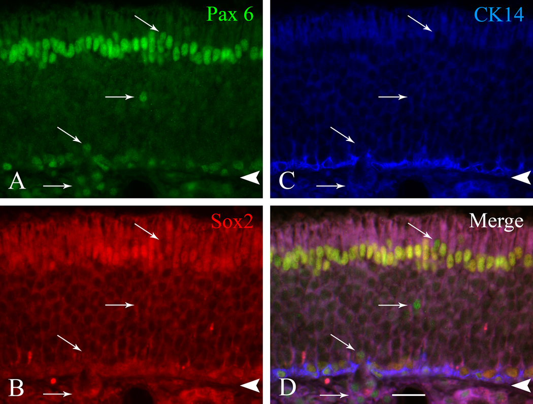
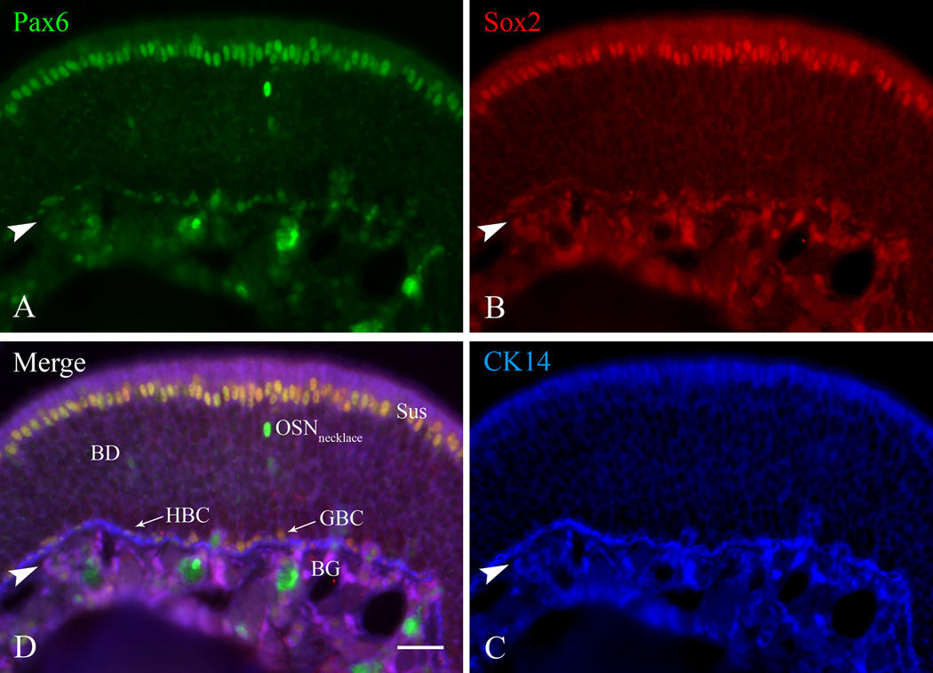
The olfactory epithelium maintains stem and progenitor cells that support the neuroepithelium's life-long capacity to reconstitute after injury. However, the identity of the stem cells--and their regulation--remain poorly defined. The transcription factors Pax6 and Sox2 are characteristic of stem cells in many tissues, including the brain. Therefore, we assessed the expression of Pax6 and Sox2 in normal olfactory epithelium and during epithelial regeneration after methyl bromide lesion or olfactory bulbectomy. Sox2 is found in multiple kinds of cells in normal epithelium, including sustentacular cells, horizontal basal cells, and some globose basal cells. Pax6 is co-expressed with Sox2 in all these, but is also found in duct/gland cells as well as olfactory neurons that innervate necklace glomeruli. Most of the Sox2/Pax6-positive globose basal cells are actively cycling, but some express the cyclin-dependent kinase inhibitor p27(Kip1), and are presumably mitotically quiescent. Among globose basal cells, Sox2 and Pax6 are co-expressed by putatively multipotent progenitors (labeled by neither anti-Mash1 nor anti-Neurog1) and neuron-committed transit amplifying cells (which express Mash1). However, Sox2 and Pax6 are expressed by only a minority of immediate neuronal precursors (Neurog1- and NeuroD1-expressing). The assignment of Sox2 and Pax6 to these categories of globose basal cells is confirmed by a temporal analysis of transcription factor expression during the recovery of the epithelium from methyl bromide-induced injury. Each of the Sox2/Pax6-colabeled cell types is at a remove from the birth of neurons; thus, suppressing their differentiation may be among the roles of Sox2/Pax6 in the olfactory epithelium.

Keywords: neural injury; neural regeneration; tissue stem cells; transcription factors.


Figures

Figure 1
Figure 1
Pax6 and Sox2 are expressed by a subgroup of GBCs. (A, C) Normal adult rat OE was stained with either anti-Pax6 antibody (in A) or anti-Sox2 antibody (in C) (both were visualized with DAB), followed by immunostaining with antibody against the horizontal basal cell (HBC) cell marker CD54 (visualized with FITC-labeled strepavidin) with corresponding insets at higher magnification in B, D, respectively. The presence of Pax6 and Sox2 in a subset of globose basal cells (GBC) is confirmed by the expression of Pax6 and the absence of CD54 in these cells, as indicated by the thick arrows. Some Pax6 (+) cells make contact with basal lamina but are not CD54 (+), as indicated by the thin arrow; basal cells such as these have also been observed with the electron microscope (Holbrook et al., 1995). BD – Bowman’s duct, BG – Bowman’s gland, both of which stain with anti-Pax6. Arrowheads mark the basal lamina. Scale bar in C is 25 µm and also applies to A. Scale bar in D is 10 µm and also applies to B.
Figure 2
Figure 2
Pax6 and Sox2 are co-expressed by the same subgroup of GBCs in rat OE. Section of normal rat OE stained with: A) anti-Pax6 (visualized with FITC-conjugated secondary antibody), B) anti-Sox2 (visualized with Texas red®-conjugated secondary antibody), C) anti-CK14 (visualized with AMCA-conjugated secondary antibody. D) Merged image. Staining with Pax6 and Sox2 is coincident and double-labeled cells appear yellow-orange in the merged image; these include Sus cells at the apex of the epithelium, HBCs marked with CK14 at the base, and GBCs situated immediately above the CK14 (+) cells. Arrows in A and D mark cells that are labeled only with Pax6 and are cells of Bowman’s gland and ducts. Arrowheads mark the basal lamina. Scale bar in D is 10 µm and also applies to the other panels.
Figure 3
Figure 3
Pax6 and Sox2 are co-expressed by the same subgroup of GBCs in the mouse OE as well. Section of normal mouse OE triple-labeled for Pax6, Sox2, and CK14. A) Anti-Pax6 staining. B) Anti-Sox2 staining. C) Anti-CK14 staining for HBCs. D) Merged image of all three labels. As in the rat OE, Pax6 labels Sus cells, Bowman’s gland (BG) and duct (BD) cells, the monolayer of HBCs apposed to the basal lamina and a population of necklace neurons (OSNnecklace) in the neuronal layers of the OE. The neuronal identity of the intensely Pax6 (+) neuronal zone cells was confirmed by staining sections of a transgenic mouse OE in which the LacZ gene is knocked into the Pax6 locus with anti-β-galactosidase and monoclonal antibody MAb213, a known necklace neuron marker (data not shown). Arrowheads mark the basal lamina. Scale bar in D is 25 µm and also applies to the other panels.
Figure 4
Figure 4
The majority of the Pax6 (+) GBCs are actively cycling. Normal rat OE stained with: A, B) anti-Pax6; C, D) anti-Ki-67, a marker of cells in all phases of the cell cycle except G0; G, H) anti-CD54, a marker of HBCs. E, F) the merged image of Pax6 and Ki67 staining. I, J) the merged image of Pax6, Ki67, and CD54. Boxed areas are shown at higher power in B, D, F, H, J. Arrows with asterisks designate Pax6 (+)/Ki-67 (+) GBCs, which are situated above the CD54 (+) HBCs. Arrows (without asterisks) mark GBCs that are only Pax6 (+) and lack detectable Ki67 labeling. Scale bar in I is 25 µm and also applies to A, C, E, G. Arrowheads mark the basal lamina. Scale bar in J is 10 µm and also applies to B, D, F, H. A magenta-green version of this figure is available online as Supplementary Figure 4.
Figure 5
Figure 5
Some Pax6 (+) GBCs express the cyclin-dependent kinase inhibitor p27Kip1 in the normal rat OE and are likely to be mitotically quiescent. Triple-labeled section of normal rat OE for Pax6, p27Kip1 and CD54. A) Anti-Pax6 staining; the arrow and arrow with asterisk designate Pax6 (+) GBCs and their surround that are shown at higher magnification in A’, A”, as indicated. B, B’, B”) Anti-p27Kip1 staining of the same section. The Pax6 (+) GBC designated by the arrow is not labeled for p27Kip1, while the GBC designated by the arrow with asterisk does express detectable p27Kip1C, C’, C”) Merged Pax6 and p27Kip1 staining, documenting that p27Kip1 and Pax6 are co-expressed in some GBCs (including the example marked by the arrow with asterisk); but most of the Pax6 (+) cells lack detectable p27Kip1 expression (including the example indicated by the solo arrow). D, D’, D”) Anti-CD54 staining to mark HBCs. E, E’, E”) The merged image of Pax6, p27Kip1 and CD54 staining to confirm that the p27Kip1 (+)/Pax6 (+) basal cells are CD54 (−) and indeed GBCs (arrow with asterisk). Arrowheads mark the basal lamina. Scale bar in E is 25 µm and also applies to A–D. Scale bar in E” is 10 µm and also applies to all other images. A magenta-green version of this figure is available online as Supplementary Figure 5.
Figure 6
Figure 6
Many Pax6 (+) GBCs are Mash1 (+). Triple-labeled section of normal rat OE for Pax6, Mash1, and CD54. Boxed areas are shown at higher magnification on the right. A, A’) Anti-Pax6 staining; Pax6 (+) GBCs are indicated by arrows (with or without asterisks). B, B’) Anti-Mash1 staining; Mash1 (+) GBCs are indicated by arrows with asterisks. C, C’) Merged image of Pax6 and Mash1 staining. D, D’) Anti- CD54 staining to mark HBCs. E, E’) Merged image of Pax6, Mash1, and CD54 staining to demonstrate that the some Pax6 (+)/Mash1 (−) basal cells are CD54 (−) and indeed GBCs. Arrowheads mark the basal lamina. Scale bar in E is 25 µm and also applies to AD. Scale bar in E’ is 10 µm and also applies to A’, D’. A magenta-green version of this figure is available online as Supplementary Figure 6.
Figure 7
Figure 7
Many Neurog1 (+) GBCs are not Pax6 (+). Section of normal rat OE double-stained with anti-Neurog1 and anti-Pax6. A, D, G, J) Anti-Neurog1 staining, a marker for GBCs that function as immediate neuronal precursors. B, E, H, K) Anti-Pax6 staining. The Neurog1 (+) GBCs indicated by the arrows are not labeled with Pax6. C, F, I, L) Merged image of Pax6 and Neurog1 staining. Neurog1 (+)-only cells in AF are indicated by arrows and are situated superficial to the Pax6 (+) GBCs. Neurog1 (+)/Pax6 (+) cells in GL are indicated by arrows with asterisks. Boxed areas are shown at higher magnification in the panels to the immediate right. Arrowheads mark the basal lamina. Scale bar in C is 25 µm and also applies to A and B. Scale bar in L is 10 µm and also applies to DK. A magenta-green version of this figure is available online as Supplementary Figure 7.
Figure 8
Figure 8
Neurog1 (+) GBCs in the OE are mainly Pax6 (−)/Sox2(−) as shown by analysis of the ΔNeurog1-eGFP BAC transgenic mouse line (generated by the Gensat project at the Rockefeller University; Gong et al., 2003). A, B) Sox2 labeling of the OE of a mouse bulbctomized 3 weeks before harvest. E, F) GFP labeling enhanced by anti-GFP staining. C, D) Merged image. Sox 2 (+) cells are found deep to the eGFP-labeled cells (arrows); the Neurog1 (+) cell marked by the arrow with asterisk is also GFP (−), but is closely apposed to GFP (+) cells. In this mouse line detectable eGFP marks the majority of Neurog1 (+) GBCs, as well as carrying over into immature neurons (see Supplementary Figure 3). Hence, most Neurog1 (+) cells in the neurogenic mouse OE do not contain detectable Sox2. Arrowheads mark the basal lamina. The scale bar in E is 25 µm and also applies to A, C. The scale bar in F is 10 µm and also applies to B, D. A magenta-green version of this figure is available online as Supplementary Figure 8.
Figure 9
Figure 9
Mash1 (+) GBCs are not p27Kip1 (+) and, thus, apparently not quiescent. Section of normal rat OE double-stained for Mash1 and p27Kip1A, A’) Anti-Mash1 staining. The Mash1 (+) GBCs are indicated by arrows, B, B’) Anti- p27Kip1 staining; the Mash1 (+) cells (arrows) are positioned deep to the layer of p27Kip1 (+) immature neurons. C, C’) Merged image of Mash1 and p27Kip1 labeling. Boxed areas are shown at higher magnification in the panels on the right. Arrowheads mark the basal lamina. Scale bar in E is 25 µm and also applies to A and E. Scale bar in F is 10 µm and also applies to B and D. A magenta-green version of this figure is available online as Supplementary Figure 9.
Figure 10
Figure 10
NeuroD1 (+) GBCs are not p27Kip1 (+) and, thus, apparently not quiescent. Section of normal rat OE double-stained for NeuroD1 and p27Kip1. In this large cluster of GBCs all of the NeuroD1 (+) cells are deep to the band of p27 labeled cells. Arrowheads mark the basal lamina. The scale bar in C is 20 µm and applies to all panels. A magenta-green version of this figure is available online as Supplementary Figure 10.
Figure 11
Figure 11
Immunolabeling for Pax6, Sox2, and Mash1 during the reconstitution of rat OE elicited by MeBr lesion. AG) Anti-Pax6 staining in unlesioned OE (Un) and at the various designated time points (1d14d) after MeBr lesion. A’G’) Anti-Sox2 staining from the same tissue at the same time points after MeBr lesion. A”G”) Anti-Mash1 staining from the same tissue at the same time points after MeBr lesion. All or the overwhelming majority of the epithelial cells are labeled with Pax6 and Sox2 at 1 and 2 days after MeBr lesion. Cells that stain with anti-Mash1 do not reappear until 2 days post lesion. The first Pax6 (−)/Sox2 (−) cells become evident at 3 days post lesion. A more prominent layer of Pax6 (−)/Sox2 (−) cells emerges and then expands from 5–14 days after lesion as neurogenesis reconstitutes the population of sensory neurons. Arrowheads mark the basal lamina. Scale bar in G” is 50 µm and applies to all other panels.
Figure 12
Figure 12
Higher magnification examination shows that all epithelial cells are Pax6 (+) and Sox2 (+) at early stages in reconstitution of the rat OE following MeBr lesion. A, B) Section from animal euthanized 1 d after lesion, double-labeled with anti-Pax6 (A) visualized with DAB and anti-CD54 (B) visualized with fluorescein-conjugated secondary antibody. Many of the cells are located superficial to the layer of CD54 (+) HBCs. C, D) Anti-Sox2 staining 1 day and 2 days after MeBr lesion visualized with DAB; all of the epithelial cells label for Sox2 at this stage. Asterisks in C designate debris that has not yet cleared from the nasal cavity after lesion. E, F) Anti-Mash1 labeling from the same tissue, showing an absence of stained cells at 1 day after lesion (E) and their reappearance at 2 days after lesion (F). Arrowheads mark the basal lamina. Scale bar in E is 10 µm and applies to all other panels.
Figure 13
Figure 13
Higher magnification examination confirms that Pax6 (−)/Sox2 (−) cells begin to re-emerge at 3 days after MeBr lesion. Triple-stained section of rat OE harvested 3 days after MeBr lesion. A) Anti-Pax6 staining – a few Pax6 (−) cells are evident at this point in the reconstitution of the OE (arrows). B) Anti-Sox2 staining – a larger number of cells are now Sox2 (−). Of these some are Pax6 (+) only (arrow with asterisk) and are presumably Bowman’s duct cells at the surface of the recovering OE. The remainder, including both GBCs and HBCs, co-express both factors and are Pax6 (+)/Sox2 (+). C) Anti-CK14 staining to mark HBCs. The Pax6 (−)/Sox2 (−) basal cells (arrows) are also CK14 (−), and hence are GBCs. Arrowheads mark the basal lamina. Scale bar in D is 10 µm and also applies to the other panels.
Figure 14
Figure 14
Expression of Sox2 and Pax6 in OE from bulbectomized rats is similar to normal, although more GBCs are labeled. AE) Anti-Sox2 staining at various time points after olfactory bulbectomy. A’–E’) Anti-Pax6 staining at various time points after olfactory bulbectomy. Note the larger population of Sox2 (+) and Pax 6 (+) basal cells at 7, 10 and 21 days after bulbectomy by comparison with the normal control. Note also the intensely stained Pax6 (+) cells within the neuronal layer of the OE at 21 days after bulbectomy, which correspond to necklace neurons. Arrowheads mark the basal lamina. Scale bar in E’ is 50 µm and also applies to the other panels.
Figure 15
Figure 15
The cells that are intensely Pax6 (+) in the neuronal zone of the OE label with a marker of, and are presumably, necklace neurons. Double-stained section of normal rat OE in a location on the ventral septum. A) Anti-Pax6 staining; the intensely positive cells situated in the neuronal layers of the OE are indicated by arrows. C) Staining with monoclonal antibody MAb213, which marks necklace neurons. Note the apical and basal processes, which resemble dendrites and axons, respectively. B) Merged image of Pax6 and MAb213 staining. Arrowheads mark the basal lamina. Scale bar in C is 25 µm and also applies to the other panels. A magenta-green version of this figure is available online as Supplementary Figure 11.

Similar articles

Cited by

References

    1. Abercrombie M. Estimation of nuclear population from microtome sections. Anat Rec. 1946;94(2):239–247. - PubMed
    1. Avilion AA, Nicolis SK, Pevny LH, Perez L, Vivian N, Lovell-Badge R. Multipotent cell lineages in early mouse development depend on SOX2 function. Genes Dev. 2003;17(1):126–140. - PMC - PubMed
    1. Baltanas FC, Weruaga E, Murias AR, Gomez C, Curto GG, Alonso JR. Chemical characterization of Pax6-immunoreactive periglomerular neurons in the mouse olfactory bulb. Cell Mol Neurobiol. 2009;29(8):1081–1085. - PMC - PubMed
    1. Bani-Yaghoub M, Tremblay RG, Lei JX, Zhang D, Zurakowski B, Sandhu JK, Smith B, Ribecco-Lutkiewicz M, Kennedy J, Walker PR, Sikorska M. Role of Sox2 in the development of the mouse neocortex. Dev Biol. 2006;295(1):52–66. - PubMed
    1. Blake JA, Thomas M, Thompson JA, White R, Ziman M. Perplexing Pax: from puzzle to paradigm. Dev Dyn. 2008;237(10):2791–2803. - PubMed

Publication types

MeSH terms